Investig Magn Reson Imaging.
2017 Mar;21(1):61-64. 10.13104/imri.2017.21.1.61.
Methanol-Induced Encephalopathy: a Case Report
- Affiliations
-
- 1Department of Radiology, Daejin Medical Center Bundang Jesaeng General Hospital, Seongnam-si, Korea. hapkitar@naver.com
- 2Department of Neurology, Daejin Medical Center Bundang Jesaeng General Hospital, Seongnam-si, Korea.
- 3Department of Internal Medicine, Daejin Medical Center Bundang Jesaeng General Hospital, Seongnam-si, Korea.
- KMID: 2376272
- DOI: http://doi.org/10.13104/imri.2017.21.1.61
Abstract
- A characteristic imaging finding in cases of methanol intoxication is putaminal necrosis, but its presence is usually not suspected due to its rarity. Methanol intoxication generally produces serious neurological symptoms that include visual disturbances and diminished consciousness, characteristically with metabolic acidosis. We reported the case of a 59-year-old man who was admitted to the hospital with diminished consciousness. Acute methanol intoxication was determined as the cause. Laboratory tests revealed high anion gap metabolic acidosis. Diffusion-weighted MRI indicated diffuse symmetric diffusion restriction lesions in the subcortical white matter of both cerebral hemispheres.
Keyword
MeSH Terms
Figure
-
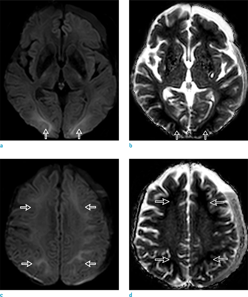
Fig. 1 59-year-old man with diminished consciousness. (a-d) Diffusion-weighted MRI shows bilateral diffuse symmetric subcortical white matter lesions with a high signal intensity and decreased apparent diffusion coefficient values (white arrows). The lesions were evenly and peripherally distributed, except for the slight predominance in both occipital lobes. These lesions did not affect the overlying cortex. There was no evidence of putaminal necrosis or hemorrhage in this patient.
Reference
-
1. Sefidbakht S, Rasekhi AR, Kamali K, et al. Methanol poisoning: acute MR and CT findings in nine patients. Neuroradiology. 2007; 49:427–435.2. Azeemuddin M, Naqi R. MRI findings in methanol intoxication: a report of three cases. J Pak Med Assoc. 2012; 62:1099–1101.3. Blanco M, Casado R, Vazquez F, Pumar JM. CT and MR imaging findings in methanol intoxication. AJNR Am J Neuroradiol. 2006; 27:452–454.4. Halavaara J, Valanne L, Setala K. Neuroimaging supports the clinical diagnosis of methanol poisoning. Neuroradiology. 2002; 44:924–928.5. Sharma P, Eesa M, Scott JN. Toxic and acquired metabolic encephalopathies: MRI appearance. AJR Am J Roentgenol. 2009; 193:879–886.6. Kraut JA, Kurtz I. Toxic alcohol ingestions: clinical features, diagnosis, and management. Clin J Am Soc Nephrol. 2008; 3:208–225.7. Osborn AG. Osborn's brain: imaging, pathology, and anatomy. 1st ed. Salt Lake City, Utah: Amirsys Pub;2013. p. 809–845.8. Longo DL, Harrison TR. Harrison's principles of internal medicine. New York, N.Y.: McGraw-Hill Medical, Cop.;2012.9. Gaul HP, Wallace CJ, Auer RN, Fong TC. MR findings in methanol intoxication. AJNR Am J Neuroradiol. 1995; 16:1783–1786.10. Ryu J, Lim KH, Ryu DR, et al. Two cases of methyl alcohol intoxication by sub-chronic inhalation and dermal exposure during aluminum CNC cutting in a small-sized subcontracted factory. Ann Occup Environ Med. 2016; 28:65.
- Full Text Links
-
- Actions
-
Cited
- CITED
-
- Close
- Share
-
- Similar articles
-
- A Case of Optic Atrophy following Methanol Poisoning
- A Case of Baclofen-induced Encephalopathy
- Letter to the Editor: A Case of Optic Nerve Atrophy with Severe Disc Cupping after Methanol Poisoning
- A Case of Bilateral Sudden Deafness Caused by Wernicke Encephalopathy
- A Case of Optic Nerve Atrophy with Severe Disc Cupping after Methanol Poisoning
