Chonnam Med J.
2017 Jan;53(1):81-82. 10.4068/cmj.2017.53.1.81.
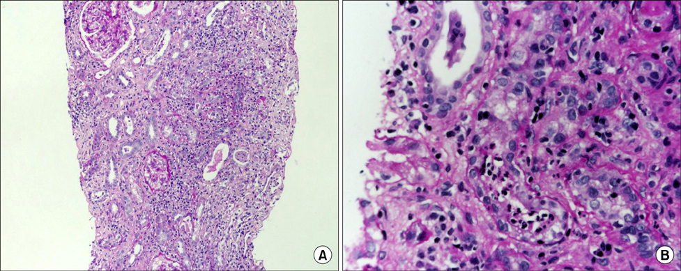
A Case of Acute Tubulointerstitial Nephritis Associated with Rifampin Therapy Presenting as Fanconi-like Syndrome
- Affiliations
-
- 1Department of Internal Medicine, Research Institute of Clinical Medicine, Chonbuk National University Medical School, Jeonju, Korea. kpkang@chonbuk.ac.kr
- KMID: 2367345
- DOI: http://doi.org/10.4068/cmj.2017.53.1.81
Abstract
- No abstract available.
MeSH Terms
Figure
Reference
-
1. Covic A, Goldsmith DJ, Segall L, Stoicescu C, Lungu S, Volovat C, et al. Rifampicin-induced acute renal failure: a series of 60 patients. Nephrol Dial Transplant. 1998; 13:924–929.
Article2. Nessi R, Bonoldi GL, Redaelli B, di Filippo G. Acute renal failure after rifampicin: a case report and survey of the literature. Nephron. 1976; 16:148–159.
Article3. Schubert C, Bates WD, Moosa MR. Acute tubulointerstitial nephritis related to antituberculous drug therapy. Clin Nephrol. 2010; 73:413–419.
Article4. Min HK, Kim EO, Lee SJ, Chang YK, Suh KS, Yang CW, et al. Rifampin-associated tubulointersititial nephritis and Fanconi syndrome presenting as hypokalemic paralysis. BMC Nephrol. 2013; 14:13.
Article
- Full Text Links
- Actions
-
Cited
- CITED
-
- Close
- Share
- Similar articles
-
- A case of Fanconi syndrome
- A Case of Tubulointerstitial Nephritis and Uveitis with Fanconi Syndrome
- An Adult Case of Tubulointerstitial Nephritis and Uveitis Syndrome in Korea
- A Case of Tubulointerstitial Nephritis and Uveitis Syndrome in an Elderly Patient
- A Case of Acute Interstitial Nephritis Complicated with Continuous Rifampin Therapy


