Spontaneous Aggressive Conversion of Venous Drainage Pattern in Dural Arteriovenous Fistula Treated with Onyx Embolization
- Affiliations
-
- 1Department of Neurosurgery, Kyung Hee University Hospital, College of Medicine, Kyung Hee University, Seoul, Korea.
- 2Department of Radiology, Kyung Hee University Hospital, College of Medicine, Kyung Hee University, Seoul, Korea. euijkim@hanmail.net
- KMID: 2367333
- DOI: http://doi.org/10.7461/jcen.2016.18.4.396
Abstract
- We report a case of dural arteriovenous fistula (DAVF) that showed spontaneous conversion of venous drainage pattern from Borden type II to type III within a four month period of follow-up. Upon admission, the patient presented with aggravated neurologic status and newly developed seizure. After admission, endovascular embolization was performed through the middle meningeal artery with Onyx®. Complete obliteration of dural arteriovenous shunt was confirmed by angiography, and the patient's clinical symptoms improved. Although most cases of DAVF show benign clinical course and conversion pattern, close follow-up is required to detect potential aggravation.
Keyword
MeSH Terms
Figure
-
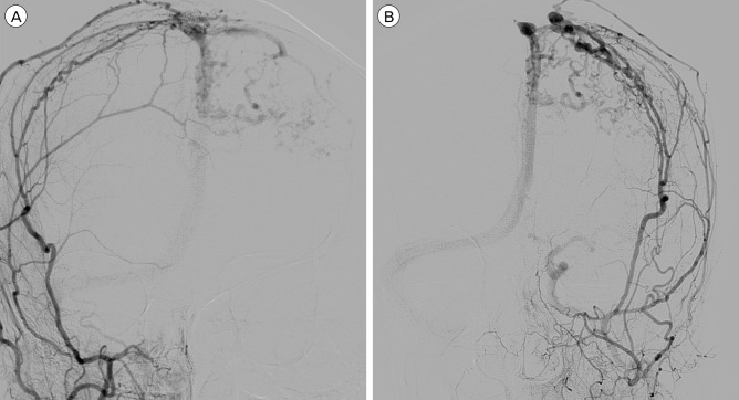
Fig. 1 Initial DSA of the patient (A : AP view of the right ECA angiography, B : AP view of the left ECA angiography) show hypervascular lesion and revealing dural arteriovenous fistula. (A) Right middle meningeal artery and superficial temporal artery supplied blood flow into superior saggital sinus via arteriovenous shunt. (B) Left middle meningeal artery supplied blood flow into superior saggital sinus via arteriovenous shunt. And there is stenosis at the proximal portion of superior saggital sinus. Cortical venous reflux is shown from superior saggital sinus to left cerebral hemisphere. DSA = digital subtraction angiography; AP = anteroposterior; ECA = external carotid artery.
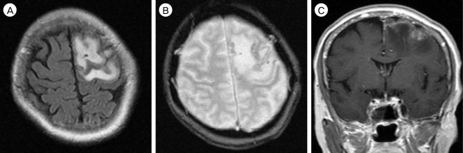
Fig. 2 Brain MRI performed when second admission after four months later from initial radiologic examination, show subacute focal infarction with minimal hemorrhagic transformation. (A) T2-weighted flair image show high signal intensity of left frontal lobe suggesting subacute cerebral infarction. (B) GRE image show multiple small low signal lesions suggesting minimal hemorrhagic transformation. (C) Mild gyral enhancement at the left frontal lobe is observed in gadolinium enhancement image. MRI = magnetic resonance image; GRE = gradient echo.
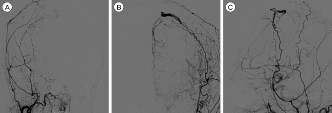
Fig. 3 Follow-up DSA after five months later from initial DSA revealing the change of venous drainage pattern. (A) Disappearance of arteriovenous shunting lesion in right ECA (external carotid artery) angiography. (B) Retrograde cortical venous reflux is shown without enhancement of superior saggital sinus in AP view of left ECA angiography. (C) There is stenotic lesion between the refluxed cortical vein and superior saggital sinus in lateral view of left ECA angiography. DSA = digital subtraction angiography; ECA = external carotid artery; AP = anteroposterior.
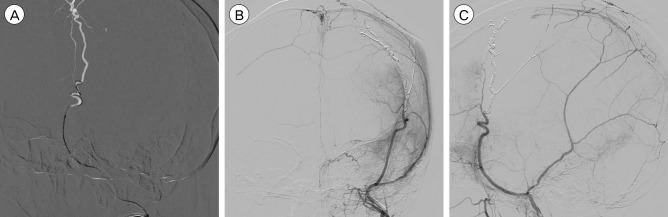
Fig. 4 After six days from second admission, Onyx embolization was performed. (A) Superselected left MMA with microcatheter and double micro-guidewire. AP view (B) and lateral view (C) after Onyx embolization show disappearance both of arteriovenous shunting lesion and cortical venous reflux. MMA = middle meningeal artery; AP = anteroposterior.
Reference
-
1. Al-Afif S, Nakamura M, Gotz F, Krauss JK. Spontaneous closure of a dural arteriovenous fistula. BMJ Case Rep. 2014; 7. 21:1–6.
Article2. Awad IA, Little JR, Akarawi WP, Ahl J. Intracranial dural arteriovenous malformations: factors predisposing to an aggressive neurological course. J Neurosurg. 1990; 6. 72(6):839–850. PMID: 2140125.
Article3. Bertalanffy A, Dietrich W, Kitz K, Bavinzski G. Treatment of dural arteriovenous fistulae (dAVF's) at the superior sagittal sinus (SSS) using embolisation combined with micro- or radiosurgery. Minim Invasive Neurosurg. 2001; 12. 44(4):205–210. PMID: 11830779.
Article4. Chaudhary MY, Sachdev VP, Cho SH, Weitzner I Jr, Puljic S, Huang YP. Dural arteriovenous malformation of the major venous sinuses: an acquired lesion. AJNR Am J Neuroradiol. 1982; Jan-Feb. 3(1):13–19. PMID: 6800236.5. Clarencon F, Biondi A, Sourour NA, Di Maria F, Iosif C, Nouet A, et al. Spontaneous closure of intracranial dural arteriovenous fistulas: a report of 3 cases. Clin Neurol Neurosurg. 2013; 7. 115(7):971–975. PMID: 23159510.6. Cognard C, Januel AC, Silva NA Jr, Tall P. Endovascular treatment of intracranial dural arteriovenous fistulas with cortical venous drainage: new management using Onyx. AJNR Am J Neuroradiol. 2008; 2. 29(2):235–241. PMID: 17989374.
Article7. Davies MA, TerBrugge K, Willinsky R, Coyne T, Saleh J, Wallace MC. The validity of classification for the clinical presentation of intracranial dural arteriovenous fistulas. J Neurosurg. 1996; 11. 85(5):830–837. PMID: 8893721.
Article8. Fermand M, Reizine D, Melki JP, Riche MC, Merland JJ. Long term follow-up of 43 pure dural arteriovenous fistulae (AVF) of the lateral sinus. Neuroradiology. 1987; 29(4):348–353. PMID: 3627416.
Article9. Ghobrial GM, Marchan E, Nair AK, Dumont AS, Tjoumakaris SI, Gonzalez LF, et al. Dural arteriovenous fistulas: a review of the literature and a presentation of a single institution's experience. World Neurosurg. 2013; Jul-Aug. 80(1-2):94–102. PMID: 22381858.
Article10. Halbach VV, Higashida RT, Hieshima GB, Rosenblum M, Cahan L. Treatment of dural arteriovenous malformations involving the superior sagittal sinus. AJNR Am J Neuroradiol. 1988; Mar-Apr. 9(2):337–343. PMID: 3128082.11. Kim DJ, terBrugge K, Krings T, Willinsky R, Wallace C. Spontaneous angiographic conversion of intracranial dural arteriovenous shunt: long-term follow-up in nontreated patients. Stroke. 2010; 7. 41(7):1489–1494. PMID: 20522815.12. Luciani A, Houdart E, Mounayer C, Saint Maurice JP, Merland JJ. Spontaneous closure of dural arteriovenous fistulas: report of three cases and review of the literature. AJNR Am J Neuroradiol. 2001; 5. 22(5):992–996. PMID: 11337347.13. Lv X, Jiang C, Zhang J, Li Y, Wu Z. Complications related to percutaneous transarterial embolization of intracranial dural arteriovenous fistulas in 40 patients. AJNR Am J Neuroradiol. 2009; 3. 30(3):462–468. PMID: 19131416.
Article14. Natarajan SK, Ghodke B, Kim LJ, Hallam DK, Britz GW, Sekhar LN. Multimodality treatment of intracranial dural arteriovenous fistulas in the Onyx era: a single center experience. World Neurosurg. 2010; 4. 73(4):365–379. PMID: 20849795.
Article15. Piske RL, Campos CM, Chaves JB, Abicalaf R, Dabus G, Batista LL, et al. Dural sinus compartment in dural arteriovenous shunts: a new angioarchitectural feature allowing superselective transvenous dural sinus occlusion treatment. AJNR Am J Neuroradiol. 2005; 8. 26(7):1715–1722. PMID: 16091520.16. Saito A, Furuno Y, Nishimura S, Kamiyama H, Nishijima M. Spontaneous closure of transverse sinus dural arteriovenous fistula: case report. Neurol Med Chir (Tokyo). 008; 12. 48(12):564–568. PMID: 19106495.17. Santillan A, Nanaszko M, Burkhardt JK, Patsalides A, Gobin YP, Riina HA. Endovascular management of intracranial dural arteriovenous fistulas: a review. Clin Neurol Neurosurg. 2013; 3. 115(3):241–251. PMID: 23287743.
Article18. Satomi J, van Dijk JM, Terbrugge KG, Willinsky RA, Wallace MC. Benign cranial dural arteriovenous fistulas: outcome of conservative management based on the natural history of the lesion. J Neurosurg. 2002; 10. 97(4):767–770. PMID: 12405361.
Article19. Soderman M, Pavic L, Edner G, Holmin S, Andersson T. Natural history of dural arteriovenous shunts. Stroke. 2008; 6. 39(6):1735–1739. PMID: 18388337.
Article20. Stiefel MF, Albuquerque FC, Park MS, Dashti SR, McDougall CG. Endovascular treatment of intracranial dural arteriovenous fistulae using Onyx: a case series. Neurosurgery. 2009; 12. 65(6 Suppl):132–139. discussion 139-40. PMID: 19934987.
Article21. Tsai LK, Jeng JS, Liu HM, Wang HJ, Yip PK. Intracranial dural arteriovenous fistulas with or without cerebral sinus thrombosis: analysis of 69 patients. J Neurol Neurosurg Psychiatry. 2004; 11. 75(11):1639–1641. PMID: 15489406.
Article22. van Dijk JM, terBrugge KG, Willinsky RA, Wallace MC. Clinical course of cranial dural arteriovenous fistulas with long-term persistent cortical venous reflux. Stroke. 2002; 5. 33(5):1233–1236. PMID: 11988596.
Article
- Full Text Links
-
- Actions
-
Cited
- CITED
-
- Close
- Share
-
- Similar articles
-
- Dural Arteriovenous Fistula Involving Transverse Sinus: Successful Embolization Using Onyx(R)
- Onyx Embolization of Dural Arteriovenous Fistula, using Scepter C Balloon Catheter: a Case Report
- Hairball-Like Migration of “Onyx Threads” into the Draining Vein during Transarterial Embolization of a Dural Arteriovenous Fistula: A Case Report and Experimental Validation
- Cortical versus Pial Venous Drainage in Dural Arteriovenous Fistula
- Dural Arteriovenous Fistula Involving an Isolated Sinus Treated Using Transarterial Onyx Embolization
