Onyx Embolization of Dural Arteriovenous Fistula, using Scepter C Balloon Catheter: a Case Report
- Affiliations
-
- 1Department of Neurosurgery, Busan Paik Hospital, School of Medicine, Inje University, Busan, Korea.
- 2Department of Diagnostic Radiology, Busan Paik Hospital, School of Medicine, Inje University, Busan, Korea. hwjeong2000@hanmail.net
- 3Department of Neurology, Busan Paik Hospital, School of Medicine, Inje University, Busan, Korea.
- KMID: 1784028
- DOI: http://doi.org/10.5469/neuroint.2013.8.2.110
Abstract
- We report our experience using Onyx for embolization of dural arteriovenous fistula (DAVF) under dual lumen balloon catheter flow arrest. Transfemoral cerebral angiography revealed a superior sagittal sinus (SSS) DAVF that was supplied via multiple branches of the external carotid arteries, the right anterior cerebral arteries, and the meningeal branches of the internal carotid artery. There was no anterograde venous drainage through the SSS, and venous drainage was almost retrograde through the medullary and cortical veins. Under general anesthesia, a transvenous approach was utilized to place the microcatheter close to the fistula site. After intravenous embolization with various coils, DAVF was partially occluded; Balloon catheter gained access to the DAVF via the right middle meningeal artery. We injected Onyx through the Scepter C catheter, after which DAVF was nearly completely occluded. Balloon-assisted Onyx embolization is a feasible and effective approach for the management of DAVF.
Keyword
MeSH Terms
-
Anesthesia, General
Anterior Cerebral Artery
Carotid Artery, External
Carotid Artery, Internal
Catheters
Central Nervous System Vascular Malformations
Cerebral Angiography
Drainage
Fistula
Hypogonadism
Meningeal Arteries
Mitochondrial Diseases
Ophthalmoplegia
Superior Sagittal Sinus
Veins
Hypogonadism
Mitochondrial Diseases
Ophthalmoplegia
Figure
-
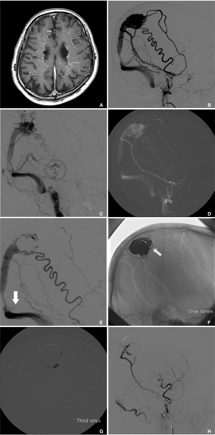
Fig. 1 A. Brain atrophy and venous congestion was observed by MRI.B. Transfemoral cerebral angiography showed the SSS DAVF was supplied via multiple branches of the external carotid artery (ECA), the right anterior cerebral arteries, and the meningeal branches of the internal carotid artery (ICA) and vertebral artery.C. Drainage through the SSS was not observed, while drainage through the vein of Labbe and sphenoparietal sinus was main (not shown). Transfemoral cerebral angiography (TFCA) revealed venous hypertension and galenic venous drainage. Furthermore, TFCA showed CVR (The hallow indicates CVR).D. Using the transvenous approach, a microcatheter (Echelon 10) was placed at the fistulous sac of the SSS.E. After intravenous coil embolization using various coils, retrograde drainage through the fistula remained (arrow).F. Scepter C balloon catheter (4×15 mm) was placed at the most distal segment of the middle meningeal artery. Tip of the balloon microcatheter was positioned at DAVF, and balloon was successfully performed (arrow).G. Over a period of 22 minutes, a total of 4.5 cc of Onyx18 was injected, and DAVF was almost completely occluded.H. After embolization, angiography revealed that no arteriovenous shunting and no cortical venous reflux occurred. Right ECA angiography.
Cited by 1 articles
-
Transvenous Onyx embolization of cavernous sinus dural arteriovenous fistula using a balloon catheter in the arterial side for flow control
Chul-Hoo Kang, Jieun Roh, Jeong A Yeom, Sang Won Lee, Seung Kug Baik
J Neurocrit Care. 2019;12(2):102-107. doi: 10.18700/jnc.190089.
Reference
-
1. Hu YC, Newman CB, Dashti SR, Albuquerque FC, McDougall CG. Cranial dural arteriovenous fistula: transarterial onyx embolization experience and technical nuances. J Neurointerv Surg. 2011; 3:5–13. PMID: 21990779.
Article2. Cognard C, Januel AC, Silva NA Jr, Tall P. Endovascular treatment of intracranial dural arteriovenous fistulas with cortical venous drainage: new management using onyx. AJNR Am J Neuroradiol. 2008; 29:235–241. PMID: 17989374.
Article3. Hoh BL, Putman CM, Budzik RF, Ogilvy CS. Surgical and endovascular flow disconnection of intracranial pial single-channel arteriovenous fistulae. Neurosurgery. 2001; 49:1351–1363. PMID: 11846934.
Article4. van Dijk JM, ter Brugge KG, Willinsky RA, Wallace MC. Clinical course of cranial dural arteriovenous fistulas with longterm persistent cortical venous reflux. Stroke. 2002; 33:1233–1236. PMID: 11988596.
Article5. Natarajan SK, Ghodke B, Kim LJ, Hallam DK, Britz GW, Sekhar LN. Multimodality treatment of intracranial dural arteriovenous fistulas in the onyx era: a single center experience. World Neurosurg. 2010; 73:365–379. PMID: 20849795.
Article6. Awad IA, Little JR, Akarawi WP, Ahl J. Intracranial dural arteriovenous malformations: factors predisposing to an aggressive neurological course. J Neurosurg. 1990; 72:839–850. PMID: 2140125.
Article7. Shi ZS, Loh Y, Duckwiler GR, Jahan R, Vinuela F. Balloon-assisted transarterial embolization of intracranial dural arteriovenous fistulas. J Neurosurg. 2009; 110:921–928. PMID: 19284242.
Article8. Fifi J, Niimi Y, Berenstein A. Onyx embolization of an extensive mandibular arteriovenous malformation via a dual lumen balloon catheter: a technical case report. J Neurointerv Surg. 2013; 5:e5. PMID: 22248630.
Article
- Full Text Links
-
- Actions
-
Cited
- CITED
-
- Close
- Share
-
- Similar articles
-
- Dural Arteriovenous Fistula Involving Transverse Sinus: Successful Embolization Using Onyx(R)
- Transarterial Balloon-assisted Onyx Embolization of Intracranial Arteriovenous Malformations Using a Dual-lumen Balloon Microcatheter: Two Case Reports
- Transvenous Onyx embolization of cavernous sinus dural arteriovenous fistula using a balloon catheter in the arterial side for flow control
- Embolization through the Ophthalmic Artery with Onyx in Bilateral Ethmoidal Dural Arteriovenous Fistula: A Case Report
- Hairball-Like Migration of “Onyx Threads” into the Draining Vein during Transarterial Embolization of a Dural Arteriovenous Fistula: A Case Report and Experimental Validation
