Neurointervention.
2023 Nov;18(3):190-194. 10.5469/neuroint.2023.00185.
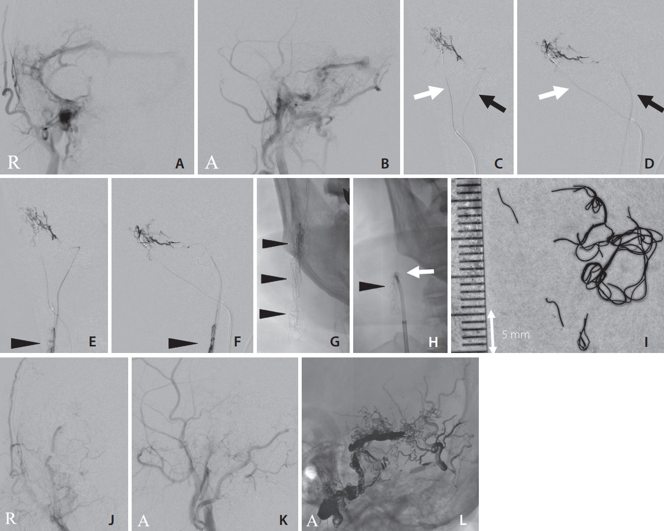
Hairball-Like Migration of “Onyx Threads” into the Draining Vein during Transarterial Embolization of a Dural Arteriovenous Fistula: A Case Report and Experimental Validation
- Affiliations
-
- 1Stroke Center, Aichi Medical University, Nagakute, Japan
- 2Neuroendovascular Therapy Center, Aichi Medical University, Nagakute, Japan
- 3Department of Neurological Surgery, Aichi Medical University, Nagakute, Japan
- KMID: 2547281
- DOI: http://doi.org/10.5469/neuroint.2023.00185
Abstract
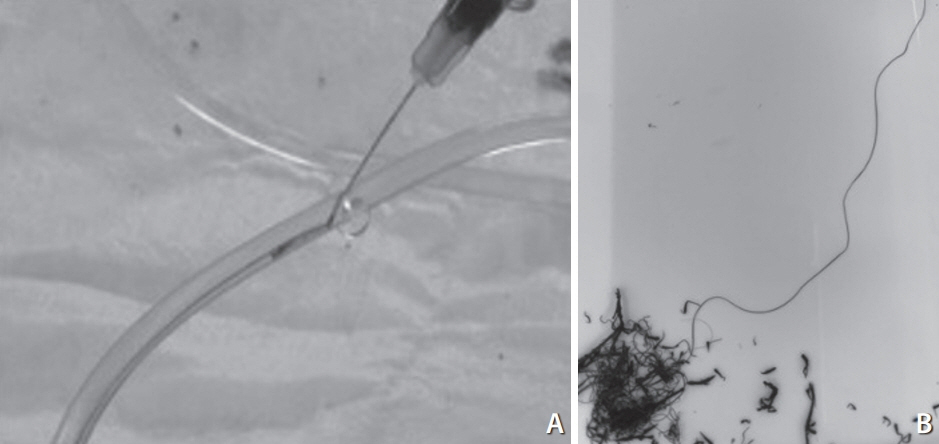
- Transarterial embolization using Onyx is a well-established treatment for dural arteriovenous fistulas (DAVFs). However, complications can arise when Onyx migrates into the venous side, impairing the draining veins. We encountered a case where Onyx, injected through the arterial side, strayed into the jugular vein, forming a hairball-like structure. Our study aimed to investigate the underlying mechanism of this unusual phenomenon. We postulated that Onyx precipitates into thread-like shapes when passing through extremely narrow openings. To test this, we extruded Onyx from a syringe through a 27-gauge needle into a silicone tube with flowing water. By varying the flow speed, we observed the hardening behavior of Onyx. Under slow flow, the extruded Onyx quickly solidified at the needle tip, forming a round mass. Conversely, high-speed flow resulted in Onyx being dispersed as small pieces. We successfully replicated the formation of “Onyx threads” under continuous slow flow conditions, similar to our case. This phenomenon occurs when Onyx unexpectedly migrates to the draining vein through a tiny opening during transarterial embolization for arteriovenous shunt diseases. Early recognition and appropriate measures are necessary to prevent occlusive complications in the draining veins and the pulmonary system.
Keyword
Figure
Reference
-
1. Mounayer C, Hammami N, Piotin M, Spelle L, Benndorf G, Kessler I, et al. Nidal embolization of brain arteriovenous malformations using Onyx in 94 patients. AJNR Am J Neuroradiol. 2007; 28:518–523.2. Nogueira RG, Dabus G, Rabinov JD, Eskey CJ, Ogilvy CS, Hirsch JA, et al. Preliminary experience with onyx embolization for the treatment of intracranial dural arteriovenous fistulas. AJNR Am J Neuroradiol. 2008; 29:91–97.
Article3. Wang H, Lv X, Jiang C, Li Y, Wu Z, Xu K. Onyx migration in the endovascular management of intracranial dural arteriovenous fistulas. Interv Neuroradiol. 2009; 15:301–308.
Article4. Piechowiak E, Zibold F, Dobrocky T, Mosimann PJ, Bervini D, Raabe A, et al. Endovascular treatment of dural arteriovenous fistulas of the transverse and sigmoid sinuses using transarterial balloon-assisted embolization combined with transvenous balloon protection of the venous sinus. AJNR Am J Neuroradiol. 2017; 38:1984–1989.
Article5. Ohshima T, Yokota M, Ogura K, Koiwai M, Matsuo N, Miyachi S. Usefulness of a curved multiple reconstruction image for transarterial intravenous coil embolization of a dural arteriovenous fistula of the sphenobasal vein: a case report. NMC Case Rep J. 2022; 9:43–47.
Article
- Full Text Links
- Actions
-
Cited
- CITED
-
- Close
- Share
- Similar articles
-
- Dural Arteriovenous Fistula Involving Transverse Sinus: Successful Embolization Using Onyx(R)
- A Case of Intraosseous Dural Arteriovenous Fistulas Involving Diploic Vein Treated with Transarterial Onyx Embolization
- Dural Arteriovenous Fistula Involving an Isolated Sinus Treated Using Transarterial Onyx Embolization
- Tentorial Dural Arteriovenous Fistula Treated Using Transarterial Onyx Embolization
- Endovascular Treatment of Dural Arteriovenous Fistulas: Single Center Experience


