Healthc Inform Res.
2016 Oct;22(4):293-298. 10.4258/hir.2016.22.4.293.
Automated Detection Algorithm of Breast Masses in Three-Dimensional Ultrasound Images
- Affiliations
-
- 1Department of Bio-medical IT Convergence Research, SW/Contents Research Laboratory, Electronics & Telecommunications Research Institute, Daejeon, Korea. jwj@etri.re.kr
- 2Medical Image Processing Team, Coreline Soft Co. Ltd., Seoul, Korea.
- 3Department of Radiology, Seoul National University Hospital, Seoul, Korea.
- KMID: 2357383
- DOI: http://doi.org/10.4258/hir.2016.22.4.293
Abstract
OBJECTIVES
We propose an automatic breast mass detection algorithm in three-dimensional (3D) ultrasound (US) images using the Hough transform technique.
METHODS
One hundred twenty-five cropped images containing 68 benign and 60 malignant masses are acquired with clinical diagnosis by an experienced radiologist. The 3D US images are masked, subsampled, contrast-adjusted, and median-filtered as preprocessing steps before the Hough transform is used. Thereafter, we perform 3D Hough transform to detect spherical hyperplanes in 3D US breast image volumes, generate Hough spheres, and sort them in the order of votes. In order to reduce the number of the false positives in the breast mass detection algorithm, the Hough sphere with a mean or grey level value of the centroid higher than the mean of the 3D US image is excluded, and the remaining Hough sphere is converted into a circumscribing parallelepiped cube as breast mass lesion candidates. Finally, we examine whether or not the generated Hough cubes were overlapping each other geometrically, and the resulting Hough cubes are suggested as detected breast mass candidates.
RESULTS
An automatic breast mass detection algorithm is applied with mass detection sensitivity of 96.1% at 0.84 false positives per case, quite comparable to the results in previous research, and we note that in the case of malignant breast mass detection, every malignant mass is detected with false positives per case at a rate of 0.62.
CONCLUSIONS
The breast mass detection efficiency of our algorithm is assessed by performing a ROC analysis.
Keyword
MeSH Terms
Figure
-
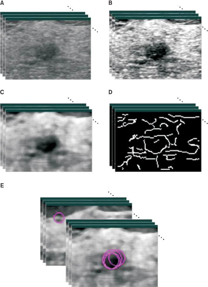
Figure 1 Illustration of pre-processing steps on 3D US images. (A) The original image. (B) RDCA is applied on (A). (C) Then, median filter is applied on (B). (D) Then, Canny edge is detected. (E) Finally, 3D Hough transform is performed on (D).
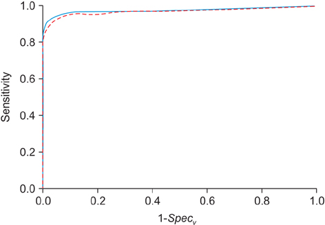
Figure 2 ROC curve of the breast mass detection algorithm using the 3D Hough transform. Solid line is for the ROC using ΔVc = 590 and dotted line for the ROC using ΔVc = 4720.
Reference
-
1. Chan HP, Wei J, Sahiner B, Rafferty EA, Wu T, Roubidoux MA, et al. Computer-aided detection system for breast masses on digital tomosynthesis mammograms: preliminary experience. Radiology. 2005; 237(3):1075–1080.
Article2. Singh S, Tourassi GD, Baker JA, Samei E, Lo JY. Automated breast mass detection in 3D reconstructed tomosynthesis volumes: a featureless approach. Med Phys. 2008; 35(8):3626–3636.
Article3. Oh WV, Kim K, Kim YJ, Kang H, Ro J, Moon W. Detection of microcalcifications in digital mammograms using foveal method. J Korean Soc Med Inform. 2009; 15(1):165–172.
Article4. Ikedo Y, Fukuoka D, Hara T, Fujita H, Takada E, Endo T, et al. Development of a fully automatic scheme for detection of masses in whole breast ultrasound images. Med Phys. 2007; 34(11):4378–4388.
Article5. Ikedo Y, Fukuoka D, Hara T, Fujita H, Takada E, Endo T, et al. Computerized mass detection in whole breast ultrasound images: reduction of false positives using bilateral subtraction technique. Proc SPIE Int Soc Opt Eng. 2007; 6514:65141.
Article6. Yu D, Lee S, Lee JW, Kim S. Automatic lesion detection and segmentation algorithm on 2D breast ultrasound images. Proc SPIE Int Soc Opt Eng. 2011; 7963:79631.
Article7. Son SH, Simonov N, Kim HJ, Lee JM, Jeon SI. Preclinical prototype development of a microwave tomography system for breast cancer detection. ETRI J. 2010; 32(6):901–910.
Article8. Chang JM, Moon WK, Cho N, Park JS, Kim SJ. Radiologists' performance in the detection of benign and malignant masses with 3D automated breast ultrasound (ABUS). Eur J Radiol. 2011; 78(1):99–103.
Article9. Chou YH, Tiu CM, Chen J, Chang RF. Automated full-field breast ultrasonography: the past and the present. J Med Ultrasound. 2007; 15(1):31–44.
Article10. Chang JM, Moon WK, Cho N, Park JS, Kim SJ. Breast cancers initially detected by hand-held ultrasound: detection performance of radiologists using automated breast ultrasound data. Acta Radiol. 2011; 52(1):8–14.
Article11. Clauser P, Londero V, Como G, Girometti R, Bazzocchi M, Zuiani C. Comparison between different imaging techniques in the evaluation of malignant breast lesions: can 3D ultrasound be useful? Radiol Med. 2014; 119(4):240–248.
Article12. Horsch K, Giger ML, Vyborny CJ, Lan L, Mendelson EB, Hendrick RE. Classification of breast lesions with multimodality computer-aided diagnosis: observer study results on an independent clinical data set. Radiology. 2006; 240(2):357–368.
Article13. Sahiner B, Chan HP, Roubidoux MA, Hadjiiski LM, Helvie MA, Paramagul C, et al. Malignant and benign breast masses on 3D US volumetric images: effect of computer-aided diagnosis on radiologist accuracy. Radiology. 2007; 242(3):716–724.
Article14. Kim JH, Cha JH, Kim N, Chang Y, Ko MS, Choi YW, et al. Computer-aided detection system for masses in automated whole breast ultrasonography: development and evaluation of the effectiveness. Ultrasonography. 2014; 33(2):105–115.
Article15. Moon WK, Lo CM, Chang JM, Huang CS, Chen JH, Chang RF. Computer-aided classification of breast masses using speckle features of automated breast ultrasound images. Med Phys. 2012; 39(10):6465–6473.
Article16. Jalalian A, Mashohor SB, Mahmud HR, Saripan MI, Ramli AR, Karasfi B. Computer-aided detection/diagnosis of breast cancer in mammography and ultrasound: a review. Clin Imaging. 2013; 37(3):420–426.
Article17. Moon WK, Shen YW, Huang CS, Chiang LR, Chang RF. Computer-aided diagnosis for the classification of breast masses in automated whole breast ultrasound images. Ultrasound Med Biol. 2011; 37(4):539–548.
Article18. Moon WK, Choi JW, Cho N, Park SH, Chang JM, Jang M, et al. Computer-aided analysis of ultrasound elasticity images for classification of benign and malignant breast masses. AJR Am J Roentgenol. 2010; 195(6):1460–1465.
Article19. Canny J. A computational approach to edge detection. IEEE Trans Pattern Anal Mach Intell. 1986; 8(6):679–698.
Article20. Yap MH, Edirisinghe EA, Bez HE. A novel algorithm for initial lesion detection in ultrasound breast images. J Appl Clin Med Phys. 2008; 9(4):2741.
Article21. Duda RO, Hart PE. Use of the Hough transformation to detect lines and curves in pictures. Commun ACM. 1972; 15(1):11–15.
Article22. Ballard DH. Generalizing the Hough transform to detect arbitrary shapes. Pattern Recognit. 1981; 13(2):111–122.
Article
- Full Text Links
-
- Actions
-
Cited
- CITED
-
- Close
- Share
-
- Similar articles
-
- A Technical Note on a Novel Technique for the Evaluation of Breast Excision Specimen: Automated Breast Ultrasound System
- Detection Rate of Breast Lesion on Mammogram Shown on Breast Sonogram
- Automated Breast Ultrasound
- Usefulness of Ultrasound-Guided Automated Core Biopsy of Nonpalpable Breast Lesions
- Clinical Application of Automated Breast Ultrasound
