Multiple Intracranial High Density Foci after Brain Parenchymal Catheterization
- Affiliations
-
- 1Department of Neurosurgery, Kwangju Christian Hospital, Gwangju, Korea. sunseinsena@hanmail.net
- KMID: 2356784
- DOI: http://doi.org/10.13004/kjnt.2016.12.2.118
Abstract
OBJECTIVE
To report an observational investigation of small high attenuated foci in computed tomography (CT) scan followed by brain parenchymal catheterization.
METHODS
From January 2011 to March 2015, we retrospectively reviewed the 381 patients who had undergone brain catheterization in our clinic and enrolled the patients who had newly developed high attenuation foci in the postoperative CT scans. The brain CT scans were reviewed about the lesion location, Hounsfield Unit (HU) and the time of appearance.
RESULTS
Twenty seven of 381 patients had high attenuation foci in CT scans after the procedure. The location of high density lesions was as follows: parenchyma in 9 (33.3%) cases, ventricle in 5 (18.5%), combined in parenchyma and ventricle in 13 (48.1%). The lesions were identified in the catheter tract in parenchymal type, and catheter-lodged frontal horn or choroid plexus in ventricular type. We could not find the calcific foci before the catheter removal, and those were found after removal in all cases. The time of appearance after the removal was variable from 0 to 14 days (mean 4.2, median 3). The regular rules of HU change in CT scans were not found as times go on.
CONCLUSION
The high attenuation foci in CT scans were bone dust originated from skull during operation. Although these lesions did not make troubles, we should clean the operation field before the insertion of brain catheter and we may use another material, like Surgicel to seal up the burr hole instead of bone dust in the end of operation.
Keyword
MeSH Terms
Figure
-
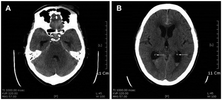
FIGURE 1 Ventricular type: (A) preoperative brain computed tomography shows aneurysmal subarachnoid hemorrhage (B) with normal calcification (white arrows) of both choroid plexus of lateral ventricle.
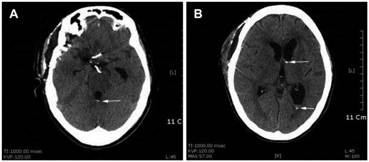
FIGURE 2 (A) unenhanced brain computed tomography on 3 days after removal of brain catheter reveals the development of hyperdense lesions (white arrows) in the 4th ventricle and (B) the left frontal and occipital horn of the lateral ventricle. (A) two aneurysmal clips are seen at Willis' circle.

FIGURE 3 Follow up computed tomography obtained 30 months after removal of catheter demonstrates that the calcific lesions are same in size and density (white arrows). (A) The two aneurysmal clips and (B) proximal catheter of ventriculo-peritoneal shunt are seen.
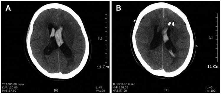
FIGURE 4 Parenchymal type: (A) preoperative unenhanced brain computed tomography (CT) shows hemorrhage in both lateral ventricles, and (B) brain CT after insertion of 2 catheters.
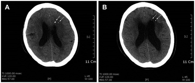
FIGURE 5 (A) unenhanced brain computed tomography on 6 days, (B) 6 months after removal of brain catheters reveal the development of hyperdense lesions (white arrows) in the left frontal lobe.
Reference
-
1. Alorainy IA. Introduction of skull bone fragments into the brain during external ventricular drain placement. Eur J Radiol. 2001; 40:218–223. PMID: 11731210.
Article2. Ji C, Ahn JG. Multiple intracranial calcifications as a complication of external ventricular drain placement. J Korean Neurosurg Soc. 2010; 47:158–160. PMID: 20224720.
Article3. Kapila A. Calcification in cerebral infarction. Radiology. 1984; 153:685–687. PMID: 6494464.
Article4. Thomson S, Tyagi AK, Chumas PD. Intracranial hypertrophic calcification complicating neuroendoscopy. Report of three cases. J Neurosurg. 2003; 98:186–189. PMID: 12546371.
- Full Text Links
-
- Actions
-
Cited
- CITED
-
- Close
- Share
-
- Similar articles
-
- Factors Related to Catheter-Induced Hemorrhage after Brain Parenchymal Catheterization
- Comparison of Ventricular Type and Parenchymal Type Intracranial Pressure (ICP) Monitoring for the Severe Traumatic Brain Injury Patients
- Influence of Alcohol on Brain Volume in Social Drinkers: Evaluation with MR-Based Intracranial-Parenchymal Ratio
- Tuberculoma Masquerading as a Meningioma: Case Report
- Associated Brain Parenchymal Abnormalities in Developmental Venous Anomalies: Evaluation with Susceptibility-weighted MR Imaging
