J Cerebrovasc Endovasc Neurosurg.
2016 Sep;18(3):286-290. 10.7461/jcen.2016.18.3.286.
Multiple Spontaneous Intracerebral Hematoma without Presenting Risk Factors
- Affiliations
-
- 1Department of Pediatric Neurosurgery, Severance Children's Hospital, Yonsei College of Medicine, Seoul, Korea. shimkyuwon@yuhs.ac
- KMID: 2355656
- DOI: http://doi.org/10.7461/jcen.2016.18.3.286
Abstract
- The incidence of intracerebral hemorrhage in those aged 45-84 years is 0.3-0.5%. In people over 80 years of age, this incidence increases 25-fold compared with that of the total population. The most common causes of spontaneous intracerebral hemorrhage in the younger population are vascular malformation, aneurysm, and overuse of drugs. In contrast, common causes in the elderly include hypertension, tumors, and coagulation disorders. Here, we present a case involving a 72-year-old male patient who, without any of these predisposing conditions, was admitted to the hospital with spontaneous intracerebral hemorrhage and showed signs of multifocal intracerebral hemorrhage during his stay. We conclude that spontaneous intracerebral hemorrhage can occur without any predisposing factors, and can lead to a patient's death. Therefore, the possibility of recurrent spontaneous intracerebral hemorrhage must be considered in patients with primary spontaneous intracerebral hemorrhage.
MeSH Terms
Figure
-
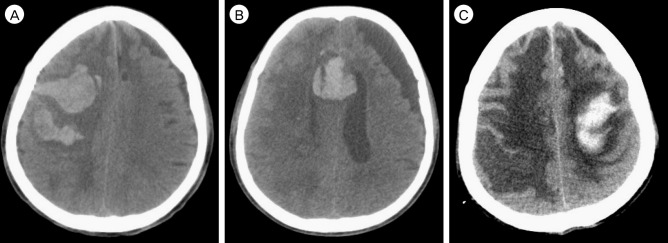
Fig. 1 CT scans of the three hemorrhages. (A) the first hemorrhage in the right frontal cortex. (B) the second hemorrhage in the septum pellucidum, which occurred 7 days after the first hemorrhage. (C) the third hemorrhage in the left fronto-parietal cortex, which occurred 17 days after the second hemorrhage. CT = computed tomography.
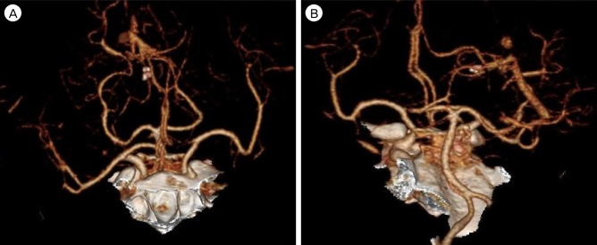
Fig. 2 Brain CT angiography revealed no specific lesions of vascular origin (A, B). CT = computed tomography.

Fig. 3 Fragmented brain tissue with hemorrhage (× 200). No other interesting outcomes were discovered.
Reference
-
1. Ariesen MJ, Claus SP, Rinkel GJ, Algra A. Risk factors for intracerebral hemorrhage in the general population: a systematic review. Stroke. 2003; 8. 34(8):2060–2065. PMID: 12843354.2. Atluru VL. Spontaneous intracerebral hematomas in juvenile diabetic ketoacidosis. Pediatr Neurol. 1986; May-Jun. 2(3):167–169. PMID: 3150280.
Article3. Bae H, Jeong D, Doh J, Lee K, Yun I, Byun B. Recurrence of bleeding in patients with hypertensive intracerebral hemorrhage. Cerebrovasc Dis. 1999; Mar-Apr. 9(2):102–108. PMID: 9973653.
Article4. Bernstein M, Fleming JF, Deck JH. Cerebral hyperperfusion after carotid endarterectomy: a cause of cerebral hemorrhage. Neurosurgery. 1984; 7. 15(1):50–56. PMID: 6472594.
Article5. Chang Y, Kargas SA, Goates JJ, Horoupian DS. Intraventricular and subarachnoid hemorrhage resulting from necrotizing vasculitis of the choroid plexus in a patient with Churg-Strauss syndrome. Clin Neuropathol. 1993; Mar-Apr. 12(2):84–87. PMID: 8097441.6. d'Angelo VA, Monte V, Scialfa G, Fiumara E, Scotti G. Intracerebral venous hemorrhage in "high-risk" carotid-cavernous fistula. Surg Neurol. 1988; 11. 30(5):387–390. PMID: 3055384.7. Fujii Y, Takeuchi S, Sasaki O, Minakawa T, Tanaka R. Multivariate analysis of predictors of hematoma enlargement in spontaneous intracerebral hemorrhage. Stroke. 1998; 6. 29(6):1160–1166. PMID: 9626289.
Article8. Guerrero MA, Schreinemakers JM, Vriens MR, Suh I, Hwang J, Shen WT. Clinical spectrum of pheochromocytoma. J Am Coll Surg. 2009; 12. 209(6):727–732. PMID: 19959041.
Article9. Hornig CR, Dorndorf W, Agnoli AL. Hemorrhagic cerebral infarction--a prospective study. Stroke. 1986; Mar-Apr. 17(2):179–185. PMID: 3515635.
Article10. Ishii N, Nishihara Y, Horie A. Amyloid angiopathy and lobar cerebral haemorrhage. J Neurol Neurosurg Psychiatry. 1984; 11. 47(11):1203–1210. PMID: 6502178.
Article11. Juvela S. Risk factors for impaired outcome after spontaneous intracerebral hemorrhage. Arch Neurol. 1995; 12. 52(12):1193–1200. PMID: 7492294.
Article12. Kumar NS, Neeraja V, Raju CG, Padala RK, Kumar TA. Multiple spontaneous hypertensive intracerebral hemorrhages. J Stroke Cerebrovasc Dis. 2015; 1. 24(1):e25–e27. PMID: 25541521.13. Lee KS, Bae HG, Yun IG. Recurrent intracerebral hemorrhage due to hypertension. Neurosurgery. 1990; 4. 26(4):586–590. PMID: 2330079.
Article14. Lee MS, Kim WC. Intracranial hemorrhage associated with idiopathic thrombocytopenic purpura: report of seven patients and a meta-analysis. Neurology. 1998; 4. 50(4):1160–1163. PMID: 9566417.
Article15. Liou HH, Liu HM, Chiang IP, Yeh TS, Chen RC. Churg-Strauss syndrome presented as multiple intracerebral hemorrhage. Lupus. 1997; 6(3):279–282. PMID: 9104737.
Article16. Mauriño J, Saposnik G, Lepera S, Rey RC, Sica RE. Multiple simultaneous intracerebral hemorrhages: clinical features and outcome. Arch Neurol. 2001; 4. 58(4):629–632. PMID: 11295994.17. Park YK, Yi HJ, Lee YJ, Cho H, Chun HJ, Oh SJ. The relationship between metabolic syndrome (MetS) and spontaneous intracerebral hemorrhage (ICH). Neurol Sci. 2013; 9. 34(9):1523–1528. PMID: 23263735.
Article18. Petri BJ, van Eijck CH, de Herder WW, Wagner A, de Krijger RR. Phaeochromocytomas and sympathetic paragangliomas. Br J Surg. 2009; 12. 96(12):1381–1392. PMID: 19918850.
Article19. Seijo M, Ucles A, Gil-Nagel A, Balseiro J, Calandre L. [Multiple cerebral hematomas: review of 7 cases]. Rev Neurol. 1996; 5. 24(129):549–553. PMID: 8681171.20. Seo JS, Nam TK, Kwon JT, Park YS. Multiple spontaneous simultaneous intracerebral hemorrhages. J Cerebrovasc Endovasc Neurosurg. 2014; 6. 16(2):104–111. PMID: 25045650.
Article21. Turner DM, Vangilder JC, Mojtahedi S, Pierson EW. Spontaneous intracerebral hematoma in carotid-cavernous fistula. Report of three cases. J Neurosurg. 1983; 10. 59(4):680–686. PMID: 6886790.
- Full Text Links
-
- Actions
-
Cited
- CITED
-
- Close
- Share
-
- Similar articles
-
- Comparative Clinical Analysis of Stereotaxic Surgery vs Conservative Treatment for Spontaneous Intracerebral Hematoma
- The Factors Affecting Enlargement of Spontaneous Intracerebral Hemorrhage
- Efficacy of Percutaneous Needle Aspiration for Evacuation of Intracerebral Hemorrhage: A Volumetric and Clincal Outcone Study
- Streotactic Evacuation and Urokinase Irrigation in the Management of Spontaneous Intracerebral Hemorrhage
- Role of 'Spot Sign' on CT Angiography to Predict Hematoma Expansion in Spontaneous Intracerebral Hemorrhage
