J Adv Prosthodont.
2016 Oct;8(5):380-387. 10.4047/jap.2016.8.5.380.
Investigation of the effects of storage time on the dimensional accuracy of impression materials using cone beam computed tomography
- Affiliations
-
- 1Department of Prosthodontics, Faculty of Dentistry, Recep Tayyip Erdogan University, Rize, Turkey. muratalkurt@hotmail.com
- 2Department of Mouth, Tooth and Jaw Radiology, Faculty of Dentistry, Inönü University, Malatya, Turkey.
- KMID: 2355442
- DOI: http://doi.org/10.4047/jap.2016.8.5.380
Abstract
- PURPOSE
The storage conditions of impressions affect the dimensional accuracy of the impression materials. The aim of the study was to assess the effects of storage time on dimensional accuracy of five different impression materials by cone beam computed tomography (CBCT).
MATERIALS AND METHODS
Polyether (Impregum), hydrocolloid (Hydrogum and Alginoplast), and silicone (Zetaflow and Honigum) impression materials were used for impressions taken from an acrylic master model. The impressions were poured and subjected to four different storage times: immediate use, and 1, 3, and 5 days of storage. Line 1 (between right and left first molar mesiobuccal cusp tips) and Line 2 (between right and left canine tips) were measured on a CBCT scanned model, and time dependent mean differences were analyzed by two-way univariate and Duncan's test (α=.05).
RESULTS
For Line 1, the total mean difference of Impregum and Hydrogum were statistically different from Alginoplast (P<.05), while Zetaflow and Honigum had smaller discrepancies. Alginoplast resulted in more difference than the other impressions (P<.05). For Line 2, the total mean difference of Impregum was statistically different from the other impressions. Significant differences were observed in Line 1 and Line 2 for the different storage periods (P<.05).
CONCLUSION
The dimensional accuracy of impression material is clinically acceptable if the impression material is stored in suitable conditions.
Keyword
Figure
-
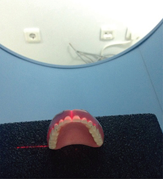
Fig. 1 Position on CBCT.

Fig. 2 (A) CBCT scan of Line 1 (between right and left first molar mesiobuccal cusp tips), (B) CBCT scan of Line 2 (between right and left canine cusp tips).

Fig. 3 Electronic caliper and the master model.

Fig. 4 Line 1 means difference of dental impression material.
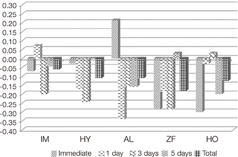
Fig. 5 Line 2 means difference of dental impression material.
Reference
-
1. Johnson GH. Impression materials. In : Craig RG, Powers JM, editors. Restorative dental materials. 11th ed. St. Louis: Elsevier;2001. p. 348–368.2. Nicholls JI. The measurement of distortion: theoretical considerations. J Prosthet Dent. 1977; 37:578–586.3. Hamalian TA, Nasr E, Chidiac JJ. Impression materials in fixed prosthodontics: influence of choice on clinical procedure. J Prosthodont. 2011; 20:153–160.4. Linke BA, Nicholls JI, Faucher RR. Distortion analysis of stone casts made from impression materials. J Prosthet Dent. 1985; 54:794–802.5. Brosky ME, Major RJ, DeLong R, Hodges JS. Evaluation of dental arch reproduction using three-dimensional optical digitization. J Prosthet Dent. 2003; 90:434–440.6. Sohmura T, Kojima T, Wakabayashi K, Takahashi J. Use of an ultrahigh-speed laser scanner for constructing three-dimensional shapes of dentition and occlusion. J Prosthet Dent. 2000; 84:345–352.7. Tarawneh FM, Panos PG, Athanasiou AE. Three-dimensional assessment of dental casts' occlusal surfaces using two impression materials. J Oral Rehabil. 2008; 35:821–826.8. Tomassetti JJ, Taloumis LJ, Denny JM, Fischer JR Jr. A comparison of 3 computerized Bolton tooth-size analyses with a commonly used method. Angle Orthod. 2001; 71:351–357.9. Elefteriadis JN, Athanasiou AE. Evaluation of impacted canines by means of computerized tomography. Int J Adult Orthodon Orthognath Surg. 1996; 11:257–264.10. Phillips RW, Ito BY. Properties of alginates. J Am Dent Assoc. 1951; 43:1–17.11. Coleman RM, Hembree JH Jr, Weber FN. Dimensional stability of irreversible hydrocolloid impression material. Am J Orthod. 1979; 75:438–446.12. Anseth KS, Bowman CN, Brannon-Peppas L. Mechanical properties of hydrogels and their experimental determination. Biomaterials. 1996; 17:1647–1657.13. Dalstra M, Melsen B. From alginate impressions to digital virtual models: accuracy and reproducibility. J Orthod. 2009; 36:36–41.14. Alcan T, Ceylanoğlu C, Baysal B. The relationship between digital model accuracy and time-dependent deformation of alginate impressions. Angle Orthod. 2009; 79:30–36.15. Sedda M, Casarotto A, Raustia A, Borracchini A. Effect of storage time on the accuracy of casts made from different irreversible hydrocolloids. J Contemp Dent Pract. 2008; 9:59–66.16. Henry PJ, Harnist DJ. Dimensional stability and accuracy of rubber impression materials. Aust Dent J. 1974; 19:162–166.17. Shellhart WC, Lange DW, Kluemper GT, Hicks EP, Kaplan AL. Reliability of the Bolton tooth-size analysis when applied to crowded dentitions. Angle Orthod. 1995; 65:327–334.18. Champagne M. Reliability of measurements from photocopies of study models. J Clin Orthod. 1992; 26:648–650.19. Lowey MN. The development of a new method of cephalometric and study cast mensuration with a computer controlled, video image capture system. Part II: Study cast mensuration. Br J Orthod. 1993; 20:315–331.20. Rossouw PE, Benatar M, Stander I, Wynchank S. A critical comparison of three methods for measuring dental models. J Dent Assoc S Afr. 1991; 46:223–226.21. Quick DC, Holtan JR, Ross GK. Use of a scanning laser three-dimensional digitizer to evaluate dimensional accuracy of dental impression materials. J Prosthet Dent. 1992; 68:229–235.22. Yamamoto K, Toshimitsu A, Mikami T, Hayashi S, Harada R, Nakamura S. Optical measurement of dental cast profile and application to analysis of three-dimensional tooth movement in orthodontics. Front Med Biol Eng. 1989; 1:119–130.23. Santoro M, Galkin S, Teredesai M, Nicolay OF, Cangialosi TJ. Comparison of measurements made on digital and plaster models. Am J Orthod Dentofacial Orthop. 2003; 124:101–105.24. Zilberman O, Huggare JA, Parikakis KA. Evaluation of the validity of tooth size and arch width measurements using conventional and three-dimensional virtual orthodontic models. Angle Orthod. 2003; 73:301–306.25. Veenema AC, Katsaros C, Boxum SC, Bronkhorst EM, Kuijpers-Jagtman AM. Index of Complexity, Outcome and Need scored on plaster and digital models. Eur J Orthod. 2009; 31:281–286.26. Bootvong K, Liu Z, McGrath C, Hägg U, Wong RW, Bendeus M, Yeung S. Virtual model analysis as an alternative approach to plaster model analysis: reliability and validity. Eur J Orthod. 2010; 32:589–595.27. Yan B, Wang L, Hu QS, Pan L, Yang K, Bao XD. Development and study of three-dimensional CT scanning system for dental cast measurement and analysis. Hua Xi Kou Qiang Yi Xue Za Zhi. 2005; 23:329–331.28. Kamegawa M, Nakamura M, Tsutsumi S. 3D morphological measurements of dental casts with occlusal relationship using microfocus X-ray CT. Dent Mater J. 2008; 27:549–554.29. Kamegawa M, Nakamura M, Kitahara K, Ohtomo H, Hasegawa T, Nakakura T, Tsutsumi S. 3D morphological assessment of occlusal treatment by measuring dental casts with a micro-focus X-ray CT. J Oral Rehabil. 2008; 35:382–389.
- Full Text Links
-
- Actions
-
Cited
- CITED
-
- Close
- Share
-
- Similar articles
-
- Three-dimensional imaging modalities in endodontics
- Evaluation of digital dental models obtained from dental cone-beam computed tomography scan of alginate impressions
- The accuracy of the imaging reformation of cone beam computed tomography for the assessment of bone defect healing
- A rare case of dilated invaginated odontome with talon cusp in a permanent maxillary central incisor diagnosed by cone beam computed tomography
- Management of root canal perforation by using cone-beam computed tomography
