Evaluation of digital dental models obtained from dental cone-beam computed tomography scan of alginate impressions
- Affiliations
-
- 1Department of Orthodontics, School of Dentistry, Chonnam National University, Gwangju, Korea. hhwang@chonnam.ac.kr
- 2Department of Orthodontics, Peking University School of Stomatology, Beijing, PR China.
- 3Department of Orthodontics, Dalian Medical University, Dalian, PR China.
- 4Dental Science Research Institute, Chonnam National University, Gwangju, Korea.
- KMID: 2164256
- DOI: http://doi.org/10.4041/kjod.2016.46.3.129
Abstract
OBJECTIVE
To investigate the dimensional accuracy of digital dental models obtained from the dental cone-beam computed tomography (CBCT) scan of alginate impressions according to the time elapse when the impressions are stored under ambient conditions.
METHODS
Alginate impressions were obtained from 20 adults using 3 different alginate materials, 2 traditional alginate materials (Alginoplast and Cavex Impressional) and 1 extended-pour alginate material (Cavex ColorChange). The impressions were stored under ambient conditions, and scanned by CBCT immediately after the impressions were taken, and then at 1 hour intervals for 6 hours. After reconstructing three-dimensional digital dental models, the models were measured and the data were analyzed to determine dimensional changes according to the elapsed time. The changes within the measurement error were regarded as clinically acceptable in this study.
RESULTS
All measurements showed a decreasing tendency with an increase in the elapsed time after the impressions. Although the extended-pour alginate exhibited a less decreasing tendency than the other 2 materials, there were no statistically significant differences between the materials. Changes above the measurement error occurred between the time points of 3 and 4 hours after the impressions.
CONCLUSIONS
The results of this study indicate that digital dental models can be obtained simply from a CBCT scan of alginate impressions without sending them to a remote laboratory. However, when the impressions are not stored under special conditions, they should be scanned immediately, or at least within 2 to 3 hours after the impressions are taken.
Keyword
Figure
-
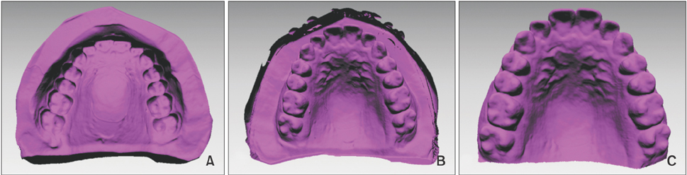
Figure 1 The process of creating a digital dental model using a cone-beam computed tomography-scanned alginate impression. A, Stereolithography file was imported into the Rapidform 2006 program (Inus, Seoul, Korea). B, The negative of the impression was converted into the positive form. C, Excessive area over the impression tray was trimmed to complete fabrication of the digital dental model.
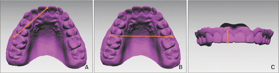
Figure 2 Linear measurements made on each digital model. A, Incisor to molar; B, intermolar width; C, incisor height.
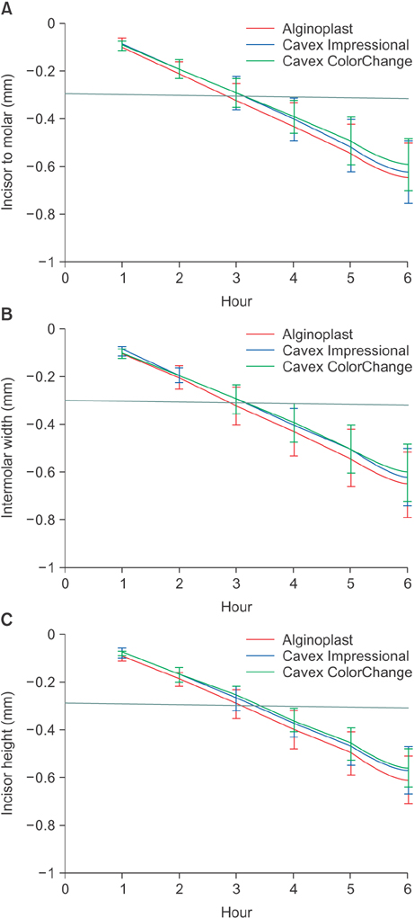
Figure 3 Graphic presentations of time-related changes and comparisons between the materials. There were no statistically significant differences between the materials although the extended-pour alginate material (Cavex ColorChange; Cavex Holland BV, Haarlem, the Netherlands) exhibited a less severe decreasing tendency than the other 2 traditional alginates. A Incisor to molar; B, intermolar width; C, incisor height.
Cited by 1 articles
-
Three-dimensional comparison of 2 digital models obtained from cone-beam computed tomographic scans of polyvinyl siloxane impressions and plaster models
Jin-Yi Park, Dasomi Kim, Sang-Sun Han, Hyung-Seog Yu, Jung-Yul Cha
Imaging Sci Dent. 2019;49(4):257-263. doi: 10.5624/isd.2019.49.4.257.
Reference
-
1. Fleming PS, Marinho V, Johal A. Orthodontic measurements on digital study models compared with plaster models: a systematic review. Orthod Craniofac Res. 2011; 14:1–16.
Article2. Naidu D, Scott J, Ong D, Ho CT. Validity, reliability and reproducibility of three methods used to measure tooth widths for bolton analyses. Aust Orthod J. 2009; 25:97–103.3. White AJ, Fallis DW, Vandewalle KS. Analysis of intra-arch and interarch measurements from digital models with 2 impression materials and a modeling process based on cone-beam computed tomography. Am J Orthod Dentofacial Orthop. 2010; 137:456.e1–456.e9.
Article4. Wiranto MG, Engelbrecht WP, Tutein Nolthenius HE, van der Meer WJ, Ren Y. Validity, reliability, and reproducibility of linear measurements on digital models obtained from intraoral and cone-beam computed tomography scans of alginate impressions. Am J Orthod Dentofacial Orthop. 2013; 143:140–147.
Article5. Naidu D, Freer TJ. Validity, reliability, and reproducibility of the iOC intraoral scanner: a comparison of tooth widths and Bolton ratios. Am J Orthod Dentofacial Orthop. 2013; 144:304–310.
Article6. Bailey E, Nelson G, Miller AJ, Andrews L, Johnson E. Predicting tooth-size discrepancy: A new formula utilizing revised landmarks and 3-dimensional laser scanning technology. Am J Orthod Dentofacial Orthop. 2013; 143:574–585.
Article7. Hayashi K, Sachdeva AU, Saitoh S, Lee SP, Kubota T, Mizoguchi I. Assessment of the accuracy and reliability of new 3-dimensional scanning devices. Am J Orthod Dentofacial Orthop. 2013; 144:619–625.
Article8. Grünheid T, McCarthy SD, Larson BE. Clinical use of a direct chairside oral scanner: an assessment of accuracy, time, and patient acceptance. Am J Orthod Dentofacial Orthop. 2014; 146:673–682.
Article9. Flügge TV, Schlager S, Nelson K, Nahles S, Metzger MC. Precision of intraoral digital dental impressions with iTero and extraoral digitization with the iTero and a model scanner. Am J Orthod Dentofacial Orthop. 2013; 144:471–478.
Article10. Patzelt SB, Lamprinos C, Stampf S, Att W. The time efficiency of intraoral scanners: an in vitro comparative study. J Am Dent Assoc. 2014; 145:542–551.11. Torassian G, Kau CH, English JD, Powers J, Bussa HI, Marie Salas-Lopez A, et al. Digital models vs plaster models using alginate and alginate substitute materials. Angle Orthod. 2010; 80:474–481.
Article12. Dahlberg G. Statistical methods for medical and biological students. London, United Kingdom: George Allen & Unwin;1940.13. Rheude B, Sadowsky PL, Ferriera A, Jacobson A. An evaluation of the use of digital study models in orthodontic diagnosis and treatment planning. Angle Orthod. 2005; 75:300–304.14. Alcan T, Ceylanoglu C, Baysal B. The relationship between digital model accuracy and time-dependent deformation of alginate impressions. Angle Orthod. 2009; 79:30–36.
Article15. Erbe C, Ruf S, Wöstmann B, Balkenhol M. Dimensional stability of contemporary irreversible hydrocolloids: humidor versus wet tissue storage. J Prosthet Dent. 2012; 108:114–122.
Article16. Walker MP, Burckhard J, Mitts DA, Williams KB. Dimensional change over time of extended-storage alginate impression materials. Angle Orthod. 2010; 80:1110–1115.
Article17. Todd JA, Oesterle LJ, Newman SM, Shellhart WC. Dimensional changes of extended-pour alginate impression materials. Am J Orthod Dentofacial Orthop. 2013; 143:4 Suppl. S55–S63.
Article
- Full Text Links
-
- Actions
-
Cited
- CITED
-
- Close
- Share
-
- Similar articles
-
- Three-dimensional comparison of 2 digital models obtained from cone-beam computed tomographic scans of polyvinyl siloxane impressions and plaster models
- Comparison of digital models generated from three-dimensional optical scanner and cone beam computed tomography
- Accuracy of Bolton analysis measured in laser scanned digital models compared with plaster models (gold standard) and cone-beam computer tomography images
- Three-dimensional imaging modalities in endodontics
- New evolution of cone-beam computed tomography in dentistry: Combining digital technologies
