Postoperative irradiation after implant placement: A pilot study for prosthetic reconstruction
- Affiliations
-
- 1Department of Advanced General Dentistry, Dankook University College of Dentistry, Cheonan, Republic of Korea.
- 2Department of Periodontology, Dental Research Institute, Seoul National University School of Dentistry, Seoul, Republic of Korea. kst72@snu.ac.kr
- 3Department of Radiation Oncology, Yonsei University College of Medicine, Seoul, Republic of Korea.
- 4Department of Prosthodontics, Yonsei University College of Dentistry, Seoul, Republic of Korea.
- 5Division in Anatomy and Development Biology, Department of Oral Biology, Yonsei University College of Dentistry, Seoul, Republic of Korea. mkchung@yuhs.ac
- 6Department of Advanced General Dentistry, Yonsei University College of Dentistry, Seoul, Republic of Korea.
- KMID: 2355440
- DOI: http://doi.org/10.4047/jap.2016.8.5.363
Abstract
- PURPOSE
On maxillofacial tumor patients, oral implant placement prior to postoperative radiotherapy can shorten the period of prosthetic reconstruction. There is still lack of research on effects of post-implant radiotherapy such as healing process or loading time, which is important for prosthodontic treatment planning. Therefore, this study evaluated the effects of post-implant local irradiation on the osseointegration of implants during different healing stages.
MATERIALS AND METHODS
Custom-made implants were placed bilaterally on maxillary posterior edentulous area 4 weeks after extraction of the maxillary first molars in Forty-eight Sprague-Dawley rats. Experimental group (exp.) received radiation after implant surgery and the other group (control) didn't. Each group was divided into three sub-groups according to the healing time (2, 4, and 8 week) from implant placement. The exp. group 1, 2 received 15-Gy radiation 1 day after implant placement (immediate irradiation). The exp. group 3 received 15-Gy radiation 4 weeks after implant placement (delayed irradiation).
RESULTS
The bone mineral density (BMD) was significantly lower in the immediate irradiation groups. BMD was similar in the delayed irradiation group and the control group. The irradiated groups exhibited a lower bone-to-implant contact ratio, although the difference was not statistically significant. The irradiated groups also exhibited a significantly lower bone volume and higher empty lacuna count than the control groups. No implant failure due to local irradiation was found in this study.
CONCLUSION
Within the limits of this study, the timing of local irradiation critically influences the bone healing mechanism, which is related to loading time of prostheses.
MeSH Terms
Figure
-
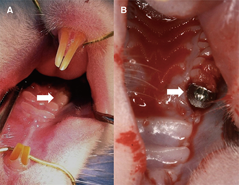
Fig. 1 Surgical procedures. (A) Maxillary first molar of the rat (white arrow). (B) Implant was placed in the first molar space after 4-week later (white arrow).
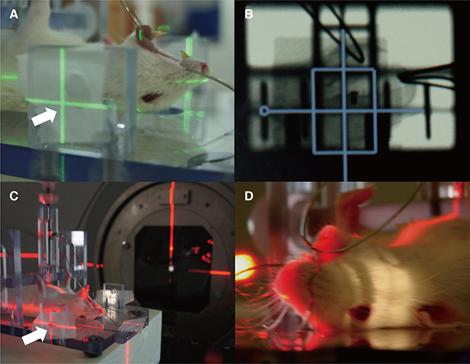
Fig. 2 Local irradiation procedures. (A, B) Verifying radiation fields using an external beam simulator (Nucletron, Veenendaal, the Netherlands). (C, D) Radiation administration using a 6.0-MV linear accelerator (Elekta, Stockholm, Sweden) to marked position (white arrow). Field size of 2 × 2 cm (see light window).
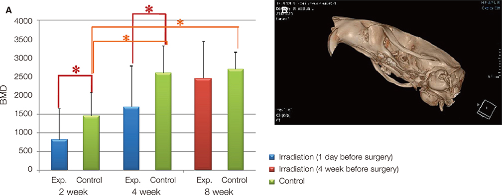
Fig. 3 Microcomputed tomography analysis. (A) Three-dimensional (3D) image reconstruction using OnDemand 3D software. (B) Bone mineral density (BMD) in the region of interest (ROI). Red stars indicate a significant difference between the exp. and control groups (P < .05); orange stars indicate a significant difference between the control groups (P < .05).
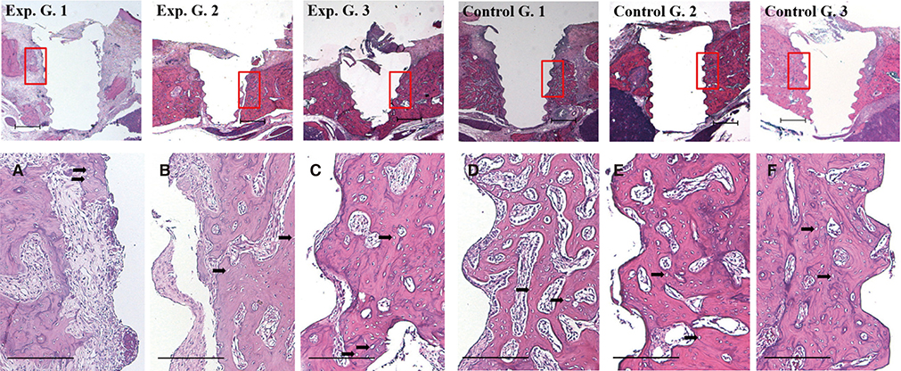
Fig. 4 Histologic images of the implant sites. Upper: Hematoxylin-and-eosin (H&E)-stained images at lower magnification (12.5×). Scale bar = 500 µm. A - C: Exp. groups. D - F: Control groups. A, D: 2 weeks after implant placement. B, E: 4 weeks after implant placement. C, F: 8 weeks after implant placement. Black arrows indicate empty lacunae (H&E, original magnification 100×. Scale bar = 200 µm).

Fig. 5 Fluorescence microscopic images of the implant sites. (A) H&E-stained image, green and red emitting regions ofexp. group 3. (B) H&E-stained image, green and red emitting regions of control group 3. Injection time of fluorescence expression agents after implant surgery are shown on the upper left (original magnification 50×. Scale bar = 200 µm).
Reference
-
1. Schepers RH, Slagter AP, Kaanders JH, van den Hoogen FJ, Merkx MA. Effect of postoperative radiotherapy on the functional result of implants placed during ablative surgery for oral cancer. Int J Oral Maxillofac Surg. 2006; 35:803–808.2. Dreizen S, Daly TE, Drane JB, Brown LR. Oral complications of cancer radiotherapy. Postgrad Med. 1977; 61:85–92.3. Granström G, Tjellström A. Effects of irradiation on osseointegration before and after implant placement: a report of three cases. Int J Oral Maxillofac Implants. 1997; 12:547–551.4. Sclaroff A, Haughey B, Gay WD, Paniello R. Immediate mandibular reconstruction and placement of dental implants. At the time of ablative surgery. Oral Surg Oral Med Oral Pathol. 1994; 78:711–717.5. Schoen PJ, Reintsema H, Raghoebar GM, Vissink A, Roodenburg JL. The use of implant retained mandibular prostheses in the oral rehabilitation of head and neck cancer patients. A review and rationale for treatment planning. Oral Oncol. 2004; 40:862–871.6. Kwakman JM, Freihofer HP, van Waas MA. Osseointegrated oral implants in head and neck cancer patients. Laryngoscope. 1997; 107:519–522.7. Brasseur M, Brogniez V, Grégoire V, Reychler H, Lengelé B, D'Hoore W, Nyssen-Behets C. Effects of irradiation on bone remodelling around mandibular implants: an experimental study in dogs. Int J Oral Maxillofac Surg. 2006; 35:850–855.8. Brogniez V, D'Hoore W, Grégoire V, Munting E, Reychler H. Implants placed in an irradiated dog mandible: a morphometric analysis. Int J Oral Maxillofac Implants. 2000; 15:511–518.9. Brogniez V, Nyssen-Behets C, Grégoire V, Reychler H, Lengelé B. Implant osseointegration in the irradiated mandible. A comparative study in dogs with a microradiographic and histologic assessment. Clin Oral Implants Res. 2002; 13:234–242.10. Schön R, Ohno K, Kudo M, Michi K. Peri-implant tissue reaction in bone irradiated the fifth day after implantation in rabbits: histologic and histomorphometric measurements. Int J Oral Maxillofac Implants. 1996; 11:228–238.11. Weinlaender M, Beumer J 3rd, Kenney EB, Lekovic V, Holmes R, Moy PK, Plenk H Jr. Histomorphometric and fluorescence microscopic evaluation of interfacial bone healing around 3 different dental implants before and after radiation therapy. Int J Oral Maxillofac Implants. 2006; 21:212–224.12. Futami T, Fujii N, Ohnishi H, Taguchi N, Kusakari H, Ohshima H, Maeda T. Tissue response to titanium implants in the rat maxilla: ultrastructural and histochemical observations of the bone-titanium interface. J Periodontol. 2000; 71:287–298.13. Del Signore A, De Sanctis V, Di Mauro E, Negri R, Perrone-Capano C, Paggi P. Gene expression pathways induced by axotomy and decentralization of rat superior cervical ganglion neurons. Eur J Neurosci. 2006; 23:65–74.14. Jegoux F, Malard O, Goyenvalle E, Aguado E, Daculsi G. Radiation effects on bone healing and reconstruction: interpretation of the literature. Oral Surg Oral Med Oral Pathol Oral Radiol Endod. 2010; 109:173–184.15. Kenzora JE, Steele RE, Yosipovitch ZH, Glimcher MJ. Experimental osteonecrosis of the femoral head in adult rabbits. Clin Orthop Relat Res. 1978; (130):8–46.16. Kim JH, Park YB, Li Z, Shim JS, Moon HS, Jung HS, Chung MK. Effect of alendronate on healing of extraction sockets and healing around implants. Oral Dis. 2011; 17:705–711.17. Andersson G, Andreasson L, Bjelkengren G. Oral implant rehabilitation in irradiated patients without adjunctive hyperbaric oxygen. Int J Oral Maxillofac Implants. 1998; 13:647–654.18. August M, Bast B, Jackson M, Perrott D. Use of the fixed mandibular implant in oral cancer patients: a retrospective study. J Oral Maxillofac Surg. 1998; 56:297–301.19. Brogniez V, Lejuste P, Pecheur A, Reychler H. Dental prosthetic reconstruction of osseointegrated implants placed in irradiated bone. Int J Oral Maxillofac Implants. 1998; 13:506–512.20. Costantino PD, Friedman CD, Steinberg MJ. Irradiated bone and its management. Otolaryngol Clin North Am. 1995; 28:1021–1038.21. Marx RE, Johnson RP. Studies in the radiobiology of osteoradionecrosis and their clinical significance. Oral Surg Oral Med Oral Pathol. 1987; 64:379–390.22. Jacobsson MG, Jönsson AK, Albrektsson TO, Turesson IE. Short- and long-term effects of irradiation on bone regeneration. Plast Reconstr Surg. 1985; 76:841–850.23. Granström G. Osseointegration in irradiated cancer patients: an analysis with respect to implant failures. J Oral Maxillofac Surg. 2005; 63:579–585.24. Jacobsson M, Albrektsson T, Turesson I. Dynamics of irradiation injury to bone tissue. A vital microscopic investigation. Acta Radiol Oncol. 1985; 24:343–350.25. Rebaudi A, Koller B, Laib A, Trisi P. Microcomputed tomographic analysis of the peri-implant bone. Int J Periodontics Restorative Dent. 2004; 24:316–325.26. Stoppie N, van der Waerden JP, Jansen JA, Duyck J, Wevers M, Naert IE. Validation of microfocus computed tomography in the evaluation of bone implant specimens. Clin Implant Dent Relat Res. 2005; 7:87–94.27. Todisco M, Trisi P. Bone mineral density and bone histomorphometry are statistically related. Int J Oral Maxillofac Implants. 2005; 20:898–904.28. Verdonck HW, Meijer GJ, Nieman FH, Stoll C, Riediger D, de Baat C. Quantitative computed tomography bone mineral density measurements in irradiated and non-irradiated minipig alveolar bone: an experimental study. Clin Oral Implants Res. 2008; 19:465–468.29. Aitasalo K. Bone tissue response to irradiation and treatment model of mandibular irradiation injury. An experimental and clinical study. Acta Otolaryngol Suppl. 1986; 428:1–54.30. Fujii N, Kusakari H, Maeda T. A histological study on tissue responses to titanium implantation in rat maxilla: the process of epithelial regeneration and bone reaction. J Periodontol. 1998; 69:485–495.31. Haga M, Fujii N, Nozawa-Inoue K, Nomura S, Oda K, Uoshima K, Maeda T. Detailed process of bone remodeling after achievement of osseointegration in a rat implantation model. Anat Rec (Hoboken). 2009; 292:38–47.32. Friedrich RE, Todorovic M, Krüll A. Simulation of scattering effects of irradiation on surroundings using the example of titanium dental implants: a Monte Carlo approach. Anticancer Res. 2010; 30:1727–1730.33. Schoen PJ, Raghoebar GM, van Oort RP, Reintsema H, van der Laan BF, Burlage FR, Roodenburg JL, Vissink A. Treatment outcome of bone-anchored craniofacial prostheses after tumor surgery. Cancer. 2001; 92:3045–3050.34. Friedrich RE, Todorovic M, Heiland M, Scheuer HA, Krüll A. Scattering effects of irradiation on surroundings calculated for a small dental implant. Anticancer Res. 2012; 32:2043–2046.35. Wang R, Pillai K, Jones PK. Dosimetric measurement of scattered radiation from dental implants in simulated head and neck radiotherapy. Int J Oral Maxillofac Implants. 1998; 13:197–203.36. Stoll P, Wächter R, Hodapp N, Schilli W. Radiation and osteosynthesis. Dosimetry on an irradiation phantom. J Craniomaxillofac Surg. 1990; 18:361–366.37. Pekmezci M, Dirican B, Yapici B, Yazici M, Alanay A, Gürdalli S. Spinal implants and radiation therapy: the effect of various configurations of titanium implant systems in a single-level vertebral metastasis model. J Bone Joint Surg Am. 2006; 88:1093–1100.38. Granström G, Jacobsson M, Tjellström A. Titanium implants in irradiated tissue: benefits from hyperbaric oxygen. Int J Oral Maxillofac Implants. 1992; 7:15–25.
- Full Text Links
-
- Actions
-
Cited
- CITED
-
- Close
- Share
-
- Similar articles
-
- A case of unexpected adjacent tooth extrusion after implant fixed prosthetic treatment, who had undergone mandibular resection and reconstruction due to ameloblastoma
- A Comparison of Permanent Expander-Implant in Immediate and Delayed Breast Reconstruction after Modified Radical Mastectomy
- Oral rehabilitation using implant supported fixed dental prostheses in a growing patient who underwent mandibulectomy and fibular free flap
- Decoronation and implant restoration of ankylosed tooth resulted from anterior avulsion: A case report
- Endoscopic Intranasal Reconstruction of Medial Orbital Wall Fracture with Muco-periosteal Flap
