Korean J Orthod.
2016 Sep;46(5):331-342. 10.4041/kjod.2016.46.5.331.
Diagnostic methods for assessing maxillary skeletal and dental transverse deficiencies: A systematic review
- Affiliations
-
- 1Department of Dentistry, School of Dentistry, University of Alberta, Edmonton, AB, Canada. carlosflores@ualberta.ca
- 2Department of Orthodontics, Case Western University, Cleveland, OH, USA.
- KMID: 2352295
- DOI: http://doi.org/10.4041/kjod.2016.46.5.331
Abstract
OBJECTIVE
To evaluate the accuracy and reliability of the diagnostic tools available for assessing maxillary transverse deficiencies.
METHODS
An electronic search of three databases was performed from their date of establishment to April 2015, with manual searching of reference lists of relevant articles. Articles were considered for inclusion if they reported the accuracy or reliability of a diagnostic method or evaluation technique for maxillary transverse dimensions in mixed or permanent dentitions. Risk of bias was assessed in the included articles, using the Quality Assessment of Diagnostic Accuracy Studies tool-2.
RESULTS
Nine articles were selected. The studies were heterogeneous, with moderate to low methodological quality, and all had a high risk of bias. Four suggested that the use of arch width prediction indices with dental cast measurements is unreliable for use in diagnosis. Frontal cephalograms derived from cone-beam computed tomography (CBCT) images were reportedly more reliable for assessing intermaxillary transverse discrepancies than posteroanterior cephalograms. Two studies proposed new three-dimensional transverse analyses with CBCT images that were reportedly reliable, but have not been validated for clinical sensitivity or specificity. No studies reported sensitivity, specificity, positive or negative predictive values or likelihood ratios, or ROC curves of the methods for the diagnosis of transverse deficiencies.
CONCLUSIONS
Current evidence does not enable solid conclusions to be drawn, owing to a lack of reliable high quality diagnostic studies evaluating maxillary transverse deficiencies. CBCT images are reportedly more reliable for diagnosis, but further validation is required to confirm CBCT's accuracy and diagnostic superiority.
Keyword
MeSH Terms
Figure
-
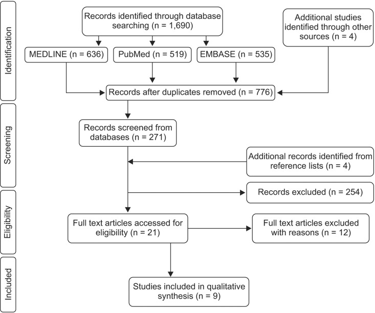
Figure 1 Flow diagram of the article selection process.
Cited by 1 articles
-
Dentofacial transverse development in Koreans according to skeletal maturation: A cross-sectional study
Soonshin Hwang, Yoonjeong Noh, Yoon Jeong Choi, Chooryung Chung, Hye Sun Lee, Kyung-Ho Kim
Korean J Orthod. 2018;48(1):39-47. doi: 10.4041/kjod.2018.48.1.39.
Reference
-
Appendix References
1. Alvaran N, Roldan SI, Buschang PH. Maxillary and mandibular arch widths of colombians. Am J Orthod Dentofacial Orthop. 2009; 135:649–656. PMID: 19409348.2. Bayome M, Park JH, Kook YA. New threedimensional cephalometric analyses among adults with a skeletal class I pattern and normal occlusion. Korean J Orthod. 2013; 43:62–73. PMID: 23671831.3. Belluzzo RH, Faltin K Jr, Ortolani C, Chelotti A. Correlation between transverse and vertical measurements in brazilian growing patients, evaluated by ricketts-faltin frontal analysis. Dental Press J Orthod. 2013; 18:50–54. PMID: 23876949.
Article4. de Oliveira MA Jr, Pereira MD, Hino CT, Campaner AB, Scanavini MA, Ferreira LM. Prediction of transverse maxillary dimension using orthodontic models. J Craniofac Surg. 2008; 19:1465–1471. PMID: 19098534.5. El-Zanaty HM, El-Beialy AR, Abou El-Ezz AM, Attia KH, El-Bialy AR, Mostafa YA. Three-dimensional dental measurements: an alternative to plaster models. Am J Orthod Dentofacial Orthop. 2010; 137:259–265. PMID: 20152684.6. Goldenberg DC, Alonso N, Goldenberg FC, Gebrin ES, Amaral TS, Scanavini MA, et al. Using computed tomography to evaluate maxillary changes after surgically assisted rapid palatal expansion. J Craniofac Surg. 2007; 18:302–311. PMID: 17414279.7. Huanca Ghislanzoni LT, Lineberger M, Cevidanes LH, Mapelli A, Sforza C, McNamara JA Jr. Evaluation of tip and torque on virtual study models: a validation study. Prog Orthod. 2013; 14:19. PMID: 24325839.
Article8. Lemieux G, Carey JP, Flores-Mir C, Secanell M, Hart A, Dietrich N, et al. Three-dimensional cephalometric superimposition of the nasomaxillary complex. Am J Orthod Dentofacial Orthop. 2014; 146:758–764. PMID: 25432257.
Article9. Ovsenik M. Assessment of malocclusion in the permanent dentition: reliability of intraoral measurements. Eur J Orthod. 2007; 29:654–659. PMID: 17962315.
Article10. Sygouros A, Motro M, Ugurlu F, Acar A. Surgically assisted rapid maxillary expansion: cone-beam computed tomography evaluation of different surgical techniques and their effects on the maxillary dentoskeletal complex. Am J Orthod Dentofacial Orthop. 2014; 146:748–757. PMID: 25432256.
Article11. Talaat S, Kaboudan A, Breuning H, Ragy N, Elshebiny T, Kula K, et al. Reliability of linear and angular dental measurements with the orthomechanics sequential analyzer. Am J Orthod Dentofacial Orthop. 2015; 147:264–269. PMID: 25636561.12. Varghese S, Kailasam V, Padmanabhan S, Vikraman B, Chithranjan A. Evaluation of the accuracy of linear measurements on spiral computed tomography-derived three-dimensional images and its comparison with digital cephalometric radiography. Dentomaxillofac Radiol. 2010; 39:216–223. PMID: 20395462.
- Full Text Links
-
- Actions
-
Cited
- CITED
-
- Close
- Share
-
- Similar articles
-
- Surgery-first Approach for Facial Asymmetry with Transverse Discrepancy Using Hyrax-type Palatal Expansion Appliance
- Cone-beam computed tomography analysis of transverse dental compensation in patients with skeletal Class III malocclusion and facial asymmetry
- Correction of unilateral posterior crossbite through the use of RME and multibracket appliances
- Dentoskeletal features in individuals with ectopic eruption of the permanent maxillary first molar
- Maxillo-mandibular Transverse Relationship of Primary Second Molar and Permanent First Molar of Children in Mixed Dentition: A Cone-Beam Computed Tomography Analysis
