Korean J Orthod.
2015 Jul;45(4):190-197. 10.4041/kjod.2015.45.4.190.
Dentoskeletal features in individuals with ectopic eruption of the permanent maxillary first molar
- Affiliations
-
- 1Department of Clinical Sciences and Traslation Medicine, University of Rome "Tor Vergata", Rome, Italy. mmucede@tin.it
- 2Private Practice, Rome, Italy.
- KMID: 2400577
- DOI: http://doi.org/10.4041/kjod.2015.45.4.190
Abstract
OBJECTIVE
The aim of the study was to analyze the prevalence and distribution of ectopic eruption of the permanent maxillary first molar (EEM) in individuals scheduled for orthodontic treatment and to investigate the association of EEM with dental characteristics, maxillary skeletal features, crowding, and other dental anomalies.
METHODS
A total of 1,317 individuals were included and randomly divided into two groups. The first 265 subjects were included as controls, while the remaining 1,052 subjects included the sample from which the final experimental EEM group was derived. The mesiodistal (M-D) crown width of the deciduous maxillary second molar and permanent maxillary first molar, maxillary arch length (A-PML), maxillomandibular transverse skeletal relationships (anterior and posterior transverse interarch discrepancies, ATID and PTID), maxillary and mandibular tooth crowding, and the presence of dental anomalies were recorded for each subject, and the statistical significance of differences in these parameters between the EEM and control groups was determined using independent sample t-tests. Chi-square tests were used to compare the prevalence of other dental anomalies between the two groups.
RESULTS
The prevalence of maxillary EEM was 2.5%. The M-D crown widths, ATID and PTID, and tooth crowding were significantly greater, while A-PML was significantly smaller, in the EEM group than in the control group. Only two subjects showed an association between EEM and maxillary lateral incisor anomalies, which included agenesis in one and microdontia in the other.
CONCLUSIONS
EEM may be a risk factor for maxillary arch constriction and severe tooth crowding.
Figure
-
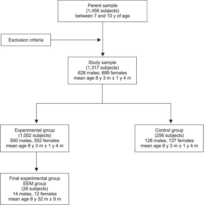
Figure 1 Study flow chart. EEM, Ectopic eruption of the permanent maxillary first molar; y, years; m, months.

Figure 2 Panoramic radiograph showing bilateral ectopic eruption of the permanent maxillary first molars in an 8-year-old subject.

Figure 3 Linear measurements on digital models. M-D crown widths, Mesiodistal crown widths; A-PML, anteroposterior maxillary length; Max-intercanine width, maxillary intercanine width; Max-intermolar width, maxillary intermolar width; Mand-intercanine width, mandibular intercanine width; Mand-intermolar width, mandibular intermolar width.
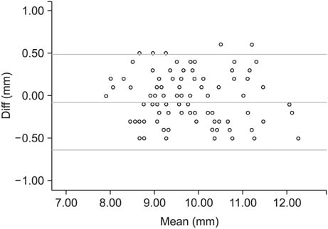
Figure 4 Results of Bland-Altman difference plot analyses. Example for M-D crown widths. Diff, difference.
Reference
-
1. Bjerklin K, Kurol J. Ectopic eruption of the maxillary first permanent molar: etiologic factors. Am J Orthod. 1983; 84:147–155.
Article2. Kurol J, Bjerklin K. Ectopic eruption of maxillary first permanent molars: a review. ASDC J Dent Child. 1986; 53:209–214.3. Bjerklin K, Kurol J. Prevalence of ectopic eruption of the maxillary first permanent molar. Swed Dent J. 1981; 5:29–34.4. Pulver F. The etiology and prevalence of ectopic eruption of the maxillary first permanent molar. ASDC J Dent Child. 1968; 35:138–146.5. Salbach A, Schremmer B, Grabowski R, Stahl de Castrillon F. Correlation between the frequency of eruption disorders for first permanent molars and the occurrence of malocclusions in early mixed dentition. J Orofac Orthop. 2012; 73:298–306.
Article6. Barberia-Leache E, Suarez-Clúa MC, Saavedra-Ontiveros D. Ectopic eruption of the maxillary first permanent molar: characteristics and occurrence in growing children. Angle Orthod. 2005; 75:610–615.7. Chintakanon K, Boonpinon P. Ectopic eruption of the first permanent molars: prevalence and etiologic factors. Angle Orthod. 1998; 68:153–160.8. Martins-Júnior PA, Marques LS. Clinical implications of early loss of a lower deciduous canine. Int J Orthod Milwaukee. 2012; 23:23–27.9. Miyamoto W, Chung CS, Yee PK. Effect of premature loss of deciduous canines and molars on malocclusion of the permanent dentition. J Dent Res. 1976; 55:584–590.
Article10. Yuen S, Chan J, Tay F. Ectopic eruption of the maxillary permanent first molar: the effect of increased mesial angulation on arch length. J Am Dent Assoc. 1985; 111:447–451.
Article11. Canut JA, Raga C. Morphological analysis of cases with ectopic eruption of the maxillary first permanent molar. Eur J Orthod. 1983; 5:249–253.
Article12. Raghoebar GM, Boering G, Vissink A, Stegenga B. Eruption disturbances of permanent molars: a review. J Oral Pathol Med. 1991; 20:159–166.
Article13. Mooney GC, Morgan AG, Rodd HD, North S. Ectopic eruption of first permanent molars: presenting features and associations. Eur Arch Paediatr Dent. 2007; 8:153–157.
Article14. Valmaseda-Castellón E, De-la-Rosa-Gay C, Gay-Escoda C. Eruption disturbances of the first and second permanent molars: results of treatment in 43 cases. Am J Orthod Dentofacial Orthop. 1999; 116:651–658.
Article15. Bjerklin K, Kurol J, Valentin J. Ectopic eruption of maxillary first permanent molars and association with other tooth and developmental disturbances. Eur J Orthod. 1992; 14:369–375.
Article16. Baccetti T. A controlled study of associated dental anomalies. Angle Orthod. 1998; 68:267–274.17. Becktor KB, Steiniche K, Kjaer I. Association between ectopic eruption of maxillary canines and first molars. Eur J Orthod. 2005; 27:186–189.
Article18. Baccetti T, Franchi L, Mc Namara JA Jr. The cervical vertebral maturation (CVM) method for the assessment of optimal treatment timing in dentofacial orthopedics. Semin Orthod. 2005; 11:119–129.
Article19. Mucedero M, Ricchiuti MR, Cozza P, Baccetti T. Prevalence rate and dentoskeletal features associated with buccally displaced maxillary canines. Eur J Orthod. 2013; 35:305–309.
Article20. Kurol J, Bjerklin K. Ectopic eruption of maxillary first permanent molars: familial tendencies. ASDC J Dent Child. 1982; 49:35–38.21. Tweed CH. Clinical orthodontics. 3th ed. St. Louis: The CV Mosby Company;1966.22. Moyers RE. Handbook of orthodontics. 4th ed. Chicago: Year Book Medical;1988.23. Tollaro I, Baccetti T, Franchi L, Tanasescu CD. Role of posterior transverse interarch discrepancy in Class II, Division 1 malocclusion during the mixed dentition phase. Am J Orthod Dentofacial Orthop. 1996; 110:417–422.
Article24. Donatelli RE, Lee SJ. How to report reliability in orthodontic research: Part 1. Am J Orthod Dentofacial Orthop. 2013; 144:156–161.
Article
- Full Text Links
-
- Actions
-
Cited
- CITED
-
- Close
- Share
-
- Similar articles
-
- A study on growth changes of maxilla and mandible and position changes of first permanent molars of growing children
- Orthodontic treatment of an eruptive disturbance of the mandibular first permanent molar
- A Case of Supernumerary Tooth within Fungus Ball in the Maxillary Sinus
- Early Eruption of Maxillary Permanent Canines : Report of 2 Cases
- Cantilever-Type Traction Appliance for Mandibular First Permanent Molars with Eruption Disturbances
