Korean J Orthod.
2016 Sep;46(5):269-279. 10.4041/kjod.2016.46.5.269.
Sphenoid bone changes in rapid maxillary expansion assessed with cone-beam computed tomography
- Affiliations
-
- 1Faculty of Medicine and Dentistry, University of Alberta, Edmonton, AB, Canada. manuel@ualberta.ca
- KMID: 2352289
- DOI: http://doi.org/10.4041/kjod.2016.46.5.269
Abstract
OBJECTIVE
Rapid maxillary expansion (RME) is used to expand the maxilla and increase arch perimeter; yet, there are few reports on its effects on the sphenoid bone. With cone-beam computed topography (CBCT), it is possible to visualize sphenoid bone changes. The purpose of this study was to investigate sphenoid bone changes observed in conjunction with RME treatments, using CBCT.
METHODS
Sixty patients (34 women and 26 men, aged 11-17 years) underwent RME as part of their orthodontic treatment. Patients were randomly assigned to one of three groups: a tooth-anchored group, a bone-anchored group, or a control group. Initial CBCT scans were performed preceding the RME treatment (Tâ‚) and again directly after the completion of expansion (Tâ‚‚). Statistical analysis included ANOVA, descriptive statistics, and the intraclass correlation coefficient (ICC).
RESULTS
The reliability of the landmark location was at least 0.783, and the largest ICC mean measurement error was 2.32 mm. With regard to distances, the largest change was 0.78 mm, which was not statistically significant (p > 0.05). Statistical significance was established in patient groups of the same sex and treatment type for the following distance measurements: right anterior lateral pterygoid plate to the right edge of the hypophyseal fossa (d₂), anterior distance between the medial pterygoid plates (d₄), and anterior distance between the left medial and lateral plates (d₈).
CONCLUSIONS
In this study, there were no clinically significant changes in the sphenoid bone due to RME treatments regardless of sex or treatment type.
MeSH Terms
Figure
-
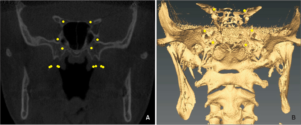
Figure 1 A frontal slice of the foramina rotundum, pterygoid canals, and optic canals in an ortho slice (A) and a volume-rendered image (B).
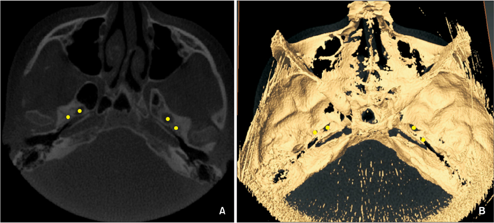
Figure 2 A coronal slice demonstrating the foramina ovale and foramina spinosum landmarks in an ortho slice (A) and a volume-rendered image (B) on AVIZO software (FEI Company, Hillsboro, OR, USA).

Figure 3 A coronal slice showing the pterygoid plate landmarks at the anterior and posterior of both the medial and lateral plates. A, Orthoslice of the landmarked structure. B, Three-dimensional view of the landmarked structure.
Reference
-
1. Angell EH. Treatment of irregularity of the permanent or adult teeth. Dent Cosmos. 1860; 1:199–600.2. Ghoneima A, Abdel-Fattah E, Hartsfield J, El-Bedwehi A, Kamel A, Kula K. Effects of rapid maxillary expansion on the cranial and circummaxillary sutures. Am J Orthod Dentofacial Orthop. 2011; 140:510–519.
Article3. Lagravère MO, Gamble J, Major PW, Heo G. Transverse dental changes after tooth-borne and bone-borne maxillary expansion. Int Orthod. 2013; 11:21–34.
Article4. Lione R, Ballanti F, Franchi L, Baccetti T, Cozza P. Treatment and posttreatment skeletal effects of rapid maxillary expansion studied with low-dose computed tomography in growing subjects. Am J Orthod Dentofacial Orthop. 2008; 134:389–392.
Article5. Timms DJ, Preston CB, Daly PF. A computed tomographic assessment of maxillary movement induced by rapid expansion - a pilot study. Eur J Orthod. 1982; 4:123–127.
Article6. Merrett SJ, Drage NA, Durning P. Cone beam computed tomography: a useful tool in orthodontic diagnosis and treatment planning. J Orthod. 2009; 36:202–210.
Article7. Gribel BF, Gribel MN, Frazäo DC, McNamara JA Jr, Manzi FR. Accuracy and reliability of craniometric measurements on lateral cephalometry and 3D measurements on CBCT scans. Angle Orthod. 2011; 81:26–35.
Article8. Leonardi R, Cutrera A, Barbato E. Rapid maxillary expansion affects the spheno-occipital synchondrosis in youngsters. A study with low-dose computed tomography. Angle Orthod. 2010; 80:106–110.
Article9. Magnusson A, Bjerklin K, Kim H, Nilsson P, Marcusson A. Three-dimensional assessment of transverse skeletal changes after surgically assisted rapid maxillary expansion and orthodontic treatment: a prospective computerized tomography study. Am J Orthod Dentofacial Orthop. 2012; 142:825–833.
Article10. Gunyuz Toklu M, Germec-Cakan D, Tozlu M. Periodontal, dentoalveolar, and skeletal effects of tooth-borne and tooth-bone-borne expansion appliances. Am J Orthod Dentofacial Orthop. 2015; 148:97–109.
Article11. Schlicher W, Nielsen I, Huang JC, Maki K, Hatcher DC, Miller AJ. Consistency and precision of landmark identification in three-dimensional cone beam computed tomography scans. Eur J Orthod. 2012; 34:263–275.
Article12. Lin L, Ahn HW, Kim SJ, Moon SC, Kim SH, Nelson G. Tooth-borne vs bone-borne rapid maxillary expanders in late adolescence. Angle Orthod. 2015; 85:253–262.
Article13. Nada RM, van Loon B, Schols JG, Maal TJ, de Koning MJ, Mostafa YA, et al. Volumetric changes of the nose and nasal airway 2 years after tooth-borne and bone-borne surgically assisted rapid maxillary expansion. Eur J Oral Sci. 2013; 121:450–456.
Article14. Vinha PP, Faria AC, Xavier SP, Christino M, de Mello-Filho FV. Enlargement of the Pharynx resulting from surgically assisted rapid maxillary expansion. J Oral Maxillofac Surg. 2016; 74:369–379.
Article15. Lima SM Jr, de Moraes M, Asprino L. Photoelastic analysis of stress distribution of surgically assisted rapid maxillary expansion with and without separation of the pterygomaxillary suture. J Oral Maxillofac Surg. 2011; 69:1771–1775.
Article16. Seeberger R, Kater W, Davids R, Thiele OC. Long term effects of surgically assisted rapid maxillary expansion without performing osteotomy of the pterygoid plates. J Craniomaxillofac Surg. 2010; 38:175–178.
Article17. Lee SC, Park JH, Bayome M, Kim KB, Araujo EA, Kook YA. Effect of bone-borne rapid maxillary expanders with and without surgical assistance on the craniofacial structures using finite element analysis. Am J Orthod Dentofacial Orthop. 2014; 145:638–648.
Article18. Bishara SE, Staley RN. Maxillary expansion: clinical implications. Am J Orthod Dentofacial Orthop. 1987; 91:3–14.
Article
- Full Text Links
-
- Actions
-
Cited
- CITED
-
- Close
- Share
-
- Similar articles
-
- A cone-beam computed tomography evaluation of buccal bone thickness following maxillary expansion
- Maxillary sinus pneumatization after maxillary molar extraction assessed with cone beam computed tomography
- Evaluation of alveolar bone loss following rapid maxillary expansion using cone-beam computed tomography
- Detection of maxillary second molar with two palatal roots using cone beam computed tomography: a case report
- Skeletal and dentoalveolar effects of different types of microimplant-assisted rapid palatal expansion
