Korean J Orthod.
2013 Apr;43(2):83-95. 10.4041/kjod.2013.43.2.83.
Evaluation of alveolar bone loss following rapid maxillary expansion using cone-beam computed tomography
- Affiliations
-
- 1Department of Orthodontics, Faculty of Dentistry, Izmir Katip Celebi University, Izmir, Turkey. tancanuysal@yahoo.com
- 2Department of Orthodontics, Faculty of Dentistry, Adnan Menderes University, Aydin, Turkey.
- 3Department of Orthodontics, Faculty of Dentistry, Dicle University, Diyarbakir, Turkey.
- 4Private Practice, Diyarbakir, Turkey.
- KMID: 1975147
- DOI: http://doi.org/10.4041/kjod.2013.43.2.83
Abstract
OBJECTIVE
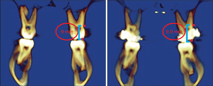
To evaluate the changes in cortical bone thickness, alveolar bone height, and the incidence of dehiscence and fenestration in the surrounding alveolar bone of posterior teeth after rapid maxillary expansion (RME) treatment using cone-beam computed tomography (CBCT).
METHODS
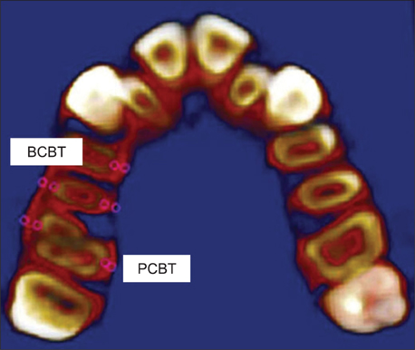
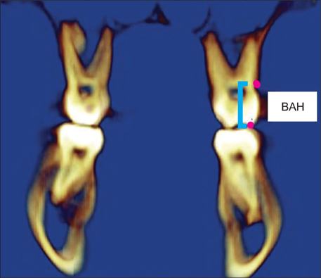
The CBCT records of 20 subjects (9 boys, mean age: 13.97 +/- 1.17 years; 11 girls, mean age: 13.53 +/- 2.12 year) that underwent RME were selected from the archives. CBCT scans had been taken before (T1) and after (T2) the RME. Moreover, 10 of the subjects had 6-month retention (T3) records. We used the CBCT data to evaluate the buccal and palatal aspects of the canines, first and second premolars, and the first molars at 3 vertical levels. The cortical bone thickness and alveolar bone height at T1 and T2 were evaluated with the paired-samples t-test or the Wilcoxon signed-rank test. Repeated measure ANOVA or the Friedman test was used to evaluate the statistical significance at T1, T2, and T3. Statistical significance was set at p < 0.05.
RESULTS
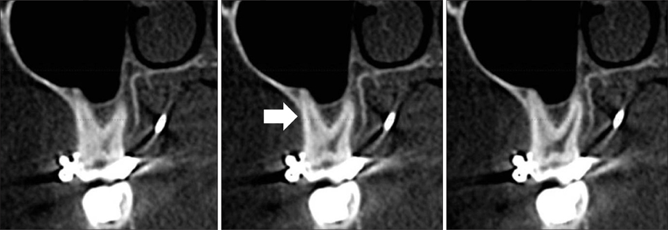
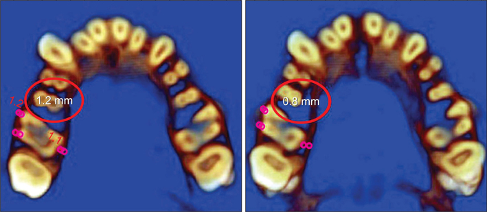
The buccal cortical bone thickness decreased gradually from baseline to the end of the retention period. After expansion, the buccal alveolar bone height was reduced significantly; however, this change was not statistically significant after the 6-month retention period. During the course of the treatment, the incidence of dehiscence and fenestration increased and decreased, respectively.
CONCLUSIONS
RME may have detrimental effects on the supporting alveolar bone, since the thickness and height of the buccal alveolar bone decreased during the retention period.
MeSH Terms
Figure
Cited by 2 articles
-
Skeletal and dentoalveolar changes after miniscrew-assisted rapid palatal expansion in young adults: A cone-beam computed tomography study
Jung Jin Park, Young-Chel Park, Kee-Joon Lee, Jung-Yul Cha, Ji Hyun Tahk, Yoon Jeong Choi
Korean J Orthod. 2017;47(2):77-86. doi: 10.4041/kjod.2017.47.2.77.Reproducibility of cone-beam computed tomographic measurements of bone plates and the interdental septum in the anterior mandible
Claudia Scigliano Valerio, Cláudia Assunção e Alves, Flávio Ricardo Manzi
Imaging Sci Dent. 2019;49(1):9-17. doi: 10.5624/isd.2019.49.1.9.
Reference
-
1. Langford SR, Sims MR. Root surface resorption, repair, and periodontal attachment following rapid maxillary expansion in man. Am J Orthod. 1982. 81:108–115.
Article2. Graber TM. Graber TM, editor. Chapter 10. Dentofacial orthopedics. Current orthodontic concepts and tech niques. 1969. Vol 11. Philadelphia: WB Saunders Company.3. Odenrick L, Karlander EL, Pierce A, Kretschmar U. Surface resorption following two forms of rapid maxillary expansion. Eur J Orthod. 1991. 13:264–270.
Article4. Rungcharassaeng K, Caruso JM, Kan JY, Kim J, Taylor G. Factors affecting buccal bone changes of maxillary posterior teeth after rapid maxillary expansion. Am J Orthod Dentofacial Orthop. 2007. 132:428.e1–428.e8.
Article5. Kartalian A, Gohl E, Adamian M, Enciso R. Cone-beam computerized tomography evaluation of the maxillary dentoskeletal complex after rapid palatal expansion. Am J Orthod Dentofacial Orthop. 2010. 138:486–492.
Article6. Garib DG, Henriques JF, Janson G, Freitas MR, Coelho RA. Rapid maxillary expansion-tooth tissue-borne versus tooth-borne expanders: a computed tomography evaluation of dentoskeletal effects. Angle Orthod. 2005. 75:548–557.7. Wainwright WM. Faciolingual tooth movement: its influence on the root and cortical plate. Am J Orthod. 1973. 64:278–302.
Article8. Baysal A, Karadede I, Hekimoglu S, Ucar F, Ozer T, Veli I, et al. Evaluation of root resorption following rapid maxillary expansion using cone-beam computed tomography. Angle Orthod. 2012. 82:488–494.
Article9. Jeffcoat MK. Current concepts in periodontal disease testing. J Am Dent Assoc. 1994. 125:1071–1078.
Article10. Molander B. Panoramic radiography in dental diagnostics. Swed Dent J Suppl. 1996. 119:1–26.11. Misch KA, Yi ES, Sarment DP. Accuracy of cone beam computed tomography for periodontal defect measurements. J Periodontol. 2006. 77:1261–1266.
Article12. Rees TD, Biggs NL, Collings CK. Radiographic interpretation of periodontal osseous lesions. Oral Surg Oral Med Oral Pathol. 1971. 32:141–153.
Article13. Hirschmann PN. Radiographic interpretation of chronic periodontitis. Int Dent J. 1987. 37:3–9.14. Vandenberghe B, Jacobs R, Yang J. Detection of periodontal bone loss using digital intraoral and cone beam computed tomography images: an in vitro assessment of bony and/or infrabony defects. Dentomaxillofac Radiol. 2008. 37:252–260.
Article15. Walker L, Enciso R, Mah J. Three-dimensional localization of maxillary canines with cone-beam computed tomography. Am J Orthod Dentofacial Orthop. 2005. 128:418–423.
Article16. Sanders DA, Rigali PH, Neace WP, Uribe F, Nanda R. Skeletal and dental asymmetries in Class II subdivision malocclusions using cone-beam computed tomography. Am J Orthod Dentofacial Orthop. 2010. 138:542.e1–542.e20.
Article17. Evangelista K, Vasconcelos Kde F, Bumann A, Hirsch E, Nitka M, Silva MA. Dehiscence and fenestration in patients with Class I and Class II Division 1 malocclusion assessed with cone-beam computed tomography. Am J Orthod Dentofacial Orthop. 2010. 138:133.e1–133.e7.
Article18. Persson RE, Hollender LG, Laurell L, Persson GR. Horizontal alveolar bone loss and vertical bone defects in an adult patient population. J Periodontol. 1998. 69:348–356.
Article19. Krebs A. Midpalatal suture expansion studies by the implant method over a seven-year period. Rep Congr Eur Orthod Soc. 1964. 40:131–142.20. Ekström C, Henrikson CO, Jensen R. Mineralization in the midpalatal suture after orthodontic expansion. Am J Orthod. 1977. 71:449–455.
Article21. Haas AJ. Rapid expansion of the maxillary dental arch and nasal cavity by opening the midpalatal suture. Angle Orthod. 1961. 31:73–90.22. Hicks EP. Slow maxillary expansion. A clinical study of the skeletal versus dental response to low-magnitude force. Am J Orthod. 1978. 73:121–141.23. Thilander B, Nyman S, Karring T, Magnusson I. Bone regeneration in alveolar bone dehiscences related to orthodontic tooth movements. Eur J Orthod. 1983. 5:105–114.
Article24. Engelking G, Zachrisson BU. Effects of incisor repositioning on monkey periodontium after expansion through the cortical plate. Am J Orthod. 1982. 82:23–32.
Article25. Barber AF, Sims MR. Rapid maxillary expansion and external root resorption in man: a scanning electron microscope study. Am J Orthod. 1981. 79:630–652.
Article26. Cotton LA. Slow maxillary expansion: skeletal versus dental response to low magnitude force in Macaca mulatta. Am J Orthod. 1978. 73:1–23.
Article27. Sarikaya S, Haydar B, Ciğer S, Ariyürek M. Changes in alveolar bone thickness due to retraction of anterior teeth. Am J Orthod Dentofacial Orthop. 2002. 122:15–26.
Article28. Leung CC, Palomo L, Griffith R, Hans MG. Accuracy and reliability of cone-beam computed tomography for measuring alveolar bone height and detecting bony dehiscences and fenestrations. Am J Orthod Dentofacial Orthop. 2010. 137:4 Suppl. S109–S119.
Article29. Fuhrmann RA, Wehrbein H, Langen HJ, Diedrich PR. Assessment of the dentate alveolar process with high resolution computed tomography. Dentomaxillofac Radiol. 1995. 24:50–54.
Article
- Full Text Links
- Actions
-
Cited
- CITED
-
- Close
- Share
- Similar articles
-
- A cone-beam computed tomography evaluation of buccal bone thickness following maxillary expansion
- Skeletal and dentoalveolar effects of different types of microimplant-assisted rapid palatal expansion
- Maxillary alveolar bone evaluation following dentoalveolar expansion with clear aligners in adults: A cone-beam computed tomography study
- Assessment of the relationship between the maxillary molars and adjacent structures using cone beam computed tomography
- Evaluation of changes in the maxillary alveolar bone after incisor intrusion

