A cone-beam computed tomography evaluation of buccal bone thickness following maxillary expansion
- Affiliations
-
- 1Department of Orthodontics, School of Dentistry, University of Texas Health Science Center at Houston, Houston, TX, USA. sercan.akyalcin@uth.tmc.edu
- 2Todd Hughes Orthodontics, Houston, TX, USA.
- KMID: 1974434
- DOI: http://doi.org/10.5624/isd.2013.43.2.85
Abstract
- PURPOSE
This study was performed to determine the buccal alveolar bone thickness following rapid maxillary expansion (RME) using cone-beam computed tomography (CBCT).
MATERIALS AND METHODS
Twenty-four individuals (15 females, 9 males; 13.9 years) that underwent RME therapy were included. Each patient had CBCT images available before (T1), after (T2), and 2 to 3 years after (T3) maxillary expansion therapy. Coronal multiplanar reconstruction images were used to measure the linear transverse dimensions, inclinations of teeth, and thickness of the buccal alveolar bone. One-way ANOVA analysis was used to compare the changes between the three times of imaging. Pairwise comparisons were made with the Bonferroni method. The level of significance was established at p<0.05.
RESULTS
The mean changes between the points in time yielded significant differences for both molar and premolar transverse measurements between T1 and T2 (p<0.05) and between T1 and T3 (p<0.05). When evaluating the effect of maxillary expansion on the amount of buccal alveolar bone, a decrease between T1 and T2 and an increase between T2 and T3 were found in the buccal bone thickness of both the maxillary first premolars and maxillary first molars. However, these changes were not significant. Similar changes were observed for the angular measurements.
CONCLUSION
RME resulted in non-significant reduction of buccal bone between T1 and T2. These changes were reversible in the long-term with no evident deleterious effects on the alveolar buccal bone.
MeSH Terms
Figure
-
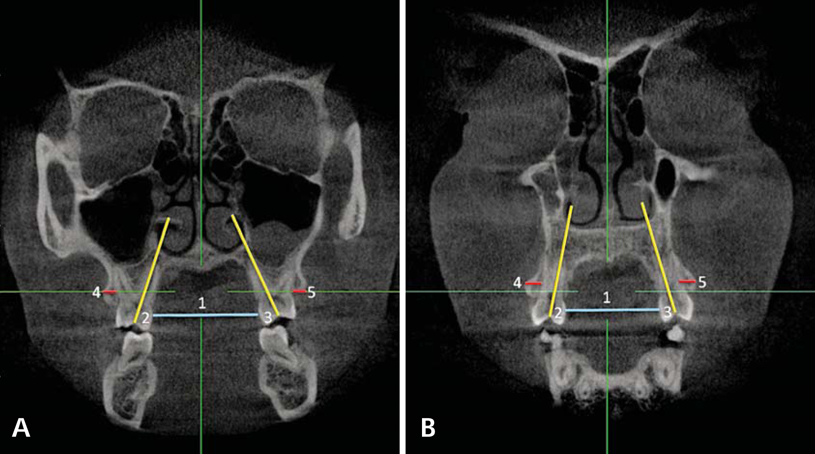
Fig. 1 Two-dimensional coronal slices perpendicular to the midsaggital plane to carry out the linear and angular measurements for this study. A. Measurements of the maxillary first molar: intermolar width (1), buccal-lingual angulation of right (2) and left (3) maxillary first molar, and right (4) and left (5) maxillary first molar buccal bone thickness. B. Measurements of the maxillary first premolar: interpremolar width (1), buccal-lingual angulation of right (2) and left (3) maxillary first premolar, and right (4) and left (5) maxillary first premolar buccal bone thickness.
Cited by 1 articles
-
Skeletal and dentoalveolar changes after miniscrew-assisted rapid palatal expansion in young adults: A cone-beam computed tomography study
Jung Jin Park, Young-Chel Park, Kee-Joon Lee, Jung-Yul Cha, Ji Hyun Tahk, Yoon Jeong Choi
Korean J Orthod. 2017;47(2):77-86. doi: 10.4041/kjod.2017.47.2.77.
Reference
-
1. Adkins MD, Nanda RS, Currier GF. Arch perimeter changes on rapid palatal expansion. Am J Orthod Dentofacial Orthop. 1990. 97:194–199.
Article2. Haas AJ. Rapid expansion of the maxillary dental arch and nasal cavity by opening the midpalatal suture. Angle Orthod. 1961. 31:73–90.3. Garib DG, Henriques JF, Janson G, de Freitas MR, Fernandes AY. Periodontal effects of rapid maxillary expansion with tooth-tissue-borne and tooth-borne expanders: a computed tomography evaluation. Am J Orthod Dentofacial Orthop. 2006. 129:749–758.
Article4. Podesser B, Williams S, Crismani AG, Bantleon HP. Evaluation of the effects of rapid maxillary expansion in growing children using computer tomography scanning: a pilot study. Eur J Orthod. 2007. 29:37–44.
Article5. Mah JK, Danforth RA, Bumann A, Hatcher D. Radiation absorbed in maxillofacial imaging with a new dental computed tomography device. Oral Surg Oral Med Oral Pathol Oral Radiol Endod. 2003. 96:508–513.
Article6. Palomo JM, Kau CH, Palomo LB, Hans MG. Three-dimensional cone beam computerized tomography in dentistry. Dent Today. 2006. 25:130–135.7. Langford SR. Root resorption extremes resulting from clinical RME. Am J Orthod. 1982. 81:371–377.
Article8. Odenrick L, Karlander EL, Pierce A, Kretschmar U. Surface resorption following two forms of rapid maxillary expansion. Eur J Orthod. 1991. 13:264–270.
Article9. Baysal A, Karadede I, Hekimoglu S, Ucar F, Ozer T, Veli I, et al. Evaluation of root resorption following rapid maxillary expansion using cone-beam computed tomography. Angle Orthod. 2012. 82:488–494.
Article10. Timock AM, Cook V, McDonald T, Leo MC, Crowe J, Benninger BL, et al. Accuracy and reliability of buccal bone height and thickness measurements from cone-beam computed tomography imaging. Am J Orthod Dentofacial Orthop. 2011. 140:734–744.
Article11. American Dental Association Council on Scientific Affairs. The use of cone-beam computed tomography in dentistry: an advisory statement from the American Dental Association Council on Scientific Affairs. J Am Dent Assoc. 2012. 143:899–902.12. Corbridge JK, Campbell PM, Taylor R, Ceen RF, Buschang PH. Transverse dentoalveolar changes after slow maxillary expansion. Am J Orthod Dentofacial Orthop. 2011. 140:317–325.
Article13. Rungcharassaeng K, Caruso JM, Kan JY, Kim J, Taylor G. Factors affecting buccal bone changes of maxillary posterior teeth after rapid maxillary expansion. Am J Orthod Dentofacial Orthop. 2007. 132:428.e1–428.e8.
Article14. Kartalian A, Gohl E, Adamian M, Enciso R. Cone-beam computerized tomography evaluation of the maxillary dentoskeletal complex after rapid palatal expansion. Am J Orthod Dentofacial Orthop. 2010. 138:486–492.
Article
- Full Text Links
-
- Actions
-
Cited
- CITED
-
- Close
- Share
-
- Similar articles
-
- Analysis of the root position and angulation of maxillary premolars in alveolar bone using cone-beam computed tomography
- Analysis of the root position of the maxillary incisors in the alveolar bone using cone-beam computed tomography
- Assessment of the relationship between the maxillary molars and adjacent structures using cone beam computed tomography
- Evaluation of alveolar bone loss following rapid maxillary expansion using cone-beam computed tomography
- Detection of maxillary second molar with two palatal roots using cone beam computed tomography: a case report
