Feasibility of Peripheral Bone Densitometry for the Assessment of Bone Density: Comparison with Dual Energy X-ray Absorptiometry of the Axial Skeleton
- Affiliations
-
- 1Department of Radiology, Kyung Hee University Medical Center, Korea. t2star@naver.com
- 2Department of Radiology, East-West Neomedical Center, Kyung Hee University, Korea.
- 3Department of Nuclear Medicine, Kyung Hee University Medical Center, Korea.
- 4Department of Preventive Medicine, School of Medicine, Kyung Hee University, Korea.
Abstract
- PURPOSE
To evaluate the feasibility of peripheral bone densitometry for the assessment of bone density, and to compare it with dual energy X-ray absorptiometry (DXA).
MATERIALS AND METHODS
Radiographic absorptiometry (RA) of the middle phalange, peripheral DXA (pDXA) of the calcaneus, and the DXA were performed for two groups: Group 1 was a normal group of 54 healthy young women and group 2 was a group of 54 postmenopausal women considered to be at a high risk for osteoporosis. For the normal group, RA and pDXA were scanned twice to assess the repeatability of the methods. The Tscores were compared to determine whether there was a correlation between the peripheral and axial bone densitometries. The cutoff values of RA and pDXA for the diagnosis of osteopenia were determined.
RESULTS
Each examination showed different T-scores for a given person. The T-scores of RA were higher than those of pDXA for the normal group, whereas the T-scores of pDXA were higher for high-risk group. The coefficients of repeatability were 0.88 in RA and 1.53 in pDXA. The correlation coefficient for DXA was higher in RA than in pDXA. The cutoff values for osteopenia were -1.773 for RA and -1.75 for pDXA, as compared to -1.0 for DXA.
CONCLUSION
The data suggests that RA is a viable screening method for osteoporosis. However, there should be consideration for the fact that bone density depends on examination methods or sites.
MeSH Terms
Figure
-
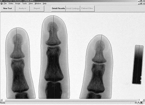
Fig. 1 Automatic identification and measurement of bone density of 2nd-4th middle phalanges, with aluminum wedge (right on figure) as a reference value.
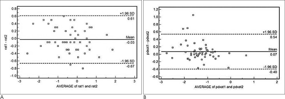
Fig. 2 Bland-Altman plot and coefficient of repeatability of the peripheral bone densitometry. A. Middle phalangeal RA (CR=0.88). B. Calcaneal pDXA (CR=1.53).
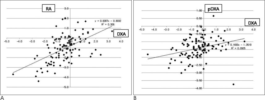
Fig. 3 Correlation between central DXA and peripheral bone densitometry. A. Lumbar DXA-middle phalangeal RA (r=0.6067). B. Lumbar DXA-calcaneal pDXA (r=0.1693).
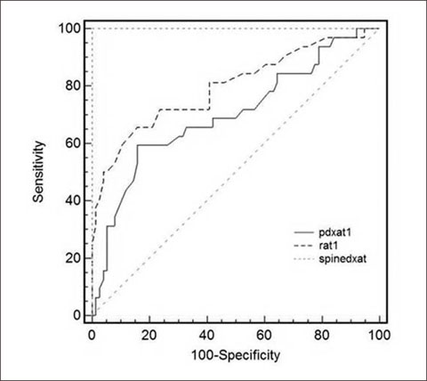
Fig. 4 Receiver operating characteristic (ROC) curves and areas under the curves for the middle phalangeal RA and calcaneal pDXA for the diagnosis of osteopenia, using a T-score=-1.0 of lumbar DXA as a standard value. pdxat1=calcaneal pDXA, rat1=middle phalangeal RA, spinedxat=lumbar DXA.
Reference
-
1. Kanis JA, Melton LJ 3rd, Christiansen C, Johnston CC, Khaltaev . The diagnosis of osteoporosis. J Bone Miner Res. 1994; 9:1137–1141.2. Huang C, Ross PD, Yates AJ, Walker RE, Imose K, Emi K, et al. Prediction of fracture risk by radiographic absorptiometry and quantitative ultrasound: a prospective study. Calcif Tissue Int. 1998; 63:380–384.3. Korean Society for Bone and Mineral Research. Diagnosis of Osteoporosis. Osteoporosis. 3rd ed. Seoul: hanmibook;2006. p. 148–159.4. Faulkner KG, von Stetten E, Miller P. Discordance in patient classification using T-scores. J Clin Densitom. 1999; 2:343–350.5. Pouillès JM, Tremollières FA, Martinez S, Delsol M, Ribot C. Ability of peripheral DXA measurements of the forearm to predict low axial bone mineral density at menopause. Osteoporosis Int. 2001; 12:71–76.6. Yang SO, Kim YS, Lee SI, Lee SH, Ham SY, Lee JH, et al. Correlation between Parameters from Quantitative Ultrasound and Bone Mineral Density by Dual x-ray Absorptiometry. J Menopausal Med. 1999; 5:452–459.7. Gasser KM, Mueller C, Zwahlen M, Kaufmann M, Fuchs G, Perrelet R, et al. Osteoporosis case finding in the general practice: phalangeal radiographic absorptiometry with and without risk factors for osteoporosis to select postmenopausal women eligible for lumbar spine and hip densitometry. Osteoporosis Int. 2005; 16:1353–1362.8. Miller PD. Bone mineral density- clinical use and application. Endocrinol Metab Clin North Am. 2003; 32:159–179.9. Leib ES, Lewiecki EM, Binkley N, Hamdy RC. Official positions of the international society for clinical densitometry. J Clin Densitom. 2004; 7:1–6.10. Kleerekoper M, Nelson DA, Flynn MJ, Pawluszka AS, Jacobsen G, Peterson EL. Comparison of radiographic absorptiometry with dual-energy X-ray absorptiometry and quantitative computed tomography in normal older white and black women. J Bone Miner Res. 1994; 9:1745–1749.11. Cummings SR, Nevitt MC, Browner WS, Stone K, Fox KM, Ensrud KE, et al. Risk factors for hip fractures in white women. N Eng J Med. 1995; 332:767–773.12. Marshall D, Johnell O, Wedel H. Meta-analysis of how well measures of bone mineral density predict occurrence of osteoporotic fractures. BMJ. 1996; 312:1254–1259.13. Kanis JA, Gluer CC. An update on the diagnosis and assessment of osteoporosis with densitometry. Osteoporosis Int. 2000; 11:192–202.14. Albanese AA, Edelson AH, Lorenze EJ, Wein EH. Quantitative radiographic survey technique for detection of bone loss. J Am Geriatr Soc. 1969; 17:142–154.15. Horsman A, Simpson M. The measurement of sequential changes in cortical bone geometry. Br J Radiol. 1975; 48:471–476.16. Michaeli DA, Mirchahi A, Singer J, Rapa FG, Plass DB, Bouxsein ML. A new X-ray based osteoporosis screening tool provides accurate and precise assessment of phalanx bone mineral content. J Clinical Densitometry. 1998; 2:23–30.17. Yang SO, Hagiwara S, Engelke K, Dhillon MS, Guglielmi G, Bendavid EJ, et al. Radiographic absorptiometry for bone mineral measurement of the phalanges: precision and accuracy study. Radiology. 1994; 192:857–859.18. Takada M, Engelke K, Hagiwara S, Grampp S, Jergas M, Gluer CC, et al. Assessment of osteoporosis: comparison of radiographic absorptiometry of the phalanges and dual X-ray absorptiometry of the radius and lumbar spine. Radiology. 1997; 202:759–763.19. Eis SR, Lewiecki EM. Peripheral bone densitometry: clinical applications. Arq Bras Endocrinol Metabol. 2006; 50:596–602.20. Miller PD, Njeh CF, Jankowski LG, Lenchik L. What are the standards by which bone mass measurement at peripheral skeletal sites should be used in the diagnosis of osteoporosis? J Clin Densitom. 2002; 5:S39–S45.21. Lenchik L, Leib ES, Hamdy RC, Binkley NC, Miller PD, Watts NB. Executive summary international society for clinical densitometry position development conference. J Clin Densitom. 2002; 5:S1–S3.
- Full Text Links
-
- Actions
-
Cited
- CITED
-
- Close
- Share
-
- Similar articles
-
- Diagnostic Value of the Bone Mineral Densitometry in the Metastatic Prostatic Cancer
- Dual-Energy X-Ray Absorptiometry: Beyond Bone Mineral Density Determination
- Measurement and Interpretation of Dual-Energy X-ray Absorptiometry Bone Density Measurements
- Clinical Application of Bone Mineral Density Measurement
- Utility of radius bone densitometry for the treatment of osteoporosis with once-weekly teriparatide therapy
