A case of inflammatory myofibroblastic tumor originated from the greater omentum in young adult
- Affiliations
-
- 1Department of Surgery, St. Vincent's Hospital, The Catholic University of Korea School of Medicine, Suwon, Korea. hmcho@catholic.ac.kr
- 2Department of Hospital Pathology, St. Vincent's Hospital, The Catholic University of Korea School of Medicine, Suwon, Korea.
Abstract
- Inflammatory myofibroblastic (IMF) tumor is a rare solid tumor that often affects children. IMF tumors occur primarily in the lung, but the tumor may affect any organ system with protean manifestations. A 22-year-old woman was evaluated for palpable low abdominal mass that had been increasing in size since two months prior. Abdominal computed tomography showed a lobulated, heterogeneous contrast enhancing soft tissue mass, 6.5 x 5.7 cm in size in the ileal mesentery. At surgery, the mass originated from the greater omentum laying in the pelvic cavity and was completely excised without tumor spillage. Histologically, the mass was a spindle cell lesion with severe atypism and some mitosis. Immunohistochemistry for anaplastic lymphoma kinase-1 revealed that the lesion was an IMF tumor. Because of its local invasiveness and its tendency to recur, this tumor can be confused with a soft tissue sarcoma. Increasing physician awareness of this entity should facilitate recognition of its clinical characteristics and laboratory findings.
MeSH Terms
Figure
-

Fig. 1 Contrast-enhanced computed tomography scan of the abdomen in axial (A) and coronal (B). A lobulated and heterogeneous contrast enhancing soft tissue mass (arrow) originating from the ileal mesentery is seen.
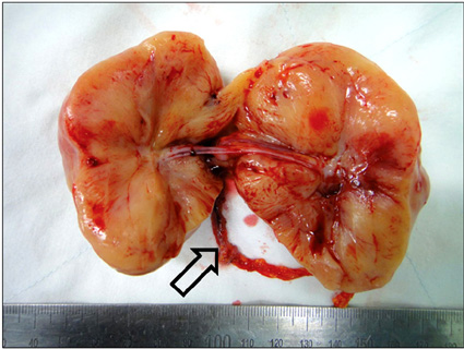
Fig. 2 Specimen consists of a portion of tan-colored tissue, measuring 7.5 × 7.0 × 5.0 cm. Arrow indicates feeding vessel originated from greater omentum.

Fig. 3 Tumor shows well-circumscribed, whorled growth pattern (H&E, ×40).
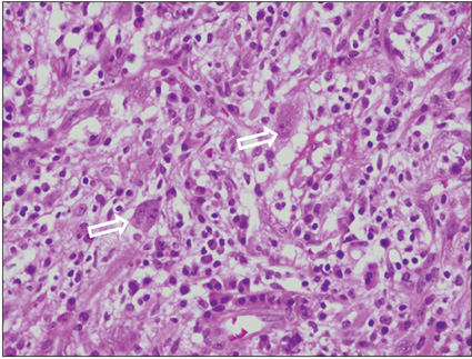
Fig. 4 Tumor is composed predominantly of spindle or stellate-shaped cells with prominent inflammatory cells. Occasionally, ganglion-like cells (arrow) present (H&E, ×400).
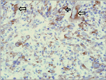
Fig. 5 Immunohistochemical staining (×400) result for anaplastic lymphoma kinase-1 shows strong positivity of tumor cytoplasm (arrow).
Reference
-
1. Coffin CM, Watterson J, Priest JR, Dehner LP. Extrapulmonary inflammatory myofibroblastic tumor (inflammatory pseudotumor): a clinicopathologic and immunohistochemical study of 84 cases. Am J Surg Pathol. 1995. 19:859–872.2. Kim SJ, Kim WS, Cheon JE, Shin SM, Youn BJ, Kim IO, et al. Inflammatory myofibroblastic tumors of the abdomen as mimickers of malignancy: imaging features in nine children. AJR Am J Roentgenol. 2009. 193:1419–1424.3. Stringer MD, Ramani P, Yeung CK, Capps SN, Kiely EM, Spitz L. Abdominal inflammatory myofibroblastic tumours in children. Br J Surg. 1992. 79:1357–1360.4. Mergan F, Jaubert F, Sauvat F, Hartmann O, Lortat-Jacob S, Revillon Y, et al. Inflammatory myofibroblastic tumor in children: clinical review with anaplastic lymphoma kinase, Epstein-Barr virus, and human herpesvirus 8 detection analysis. J Pediatr Surg. 2005. 40:1581–1586.5. Sawant S, Kasturi L, Amin A. Inflammatory myofibroblastic tumor. Indian J Pediatr. 2002. 69:1001–1002.6. Pecorella I, Ciardi A, Memeo L, Trombetta G, de Quarto A, de Simone P, et al. Inflammatory pseudotumour of the liver: evidence for malignant transformation. Pathol Res Pract. 1999. 195:115–120.7. Sodhi KS, Virmani V, Bal A, Saxena AK, Samujh R, Khandelwa N. Inflammatory pseudotumor of the omentum. Indian J Pediatr. 2010. 77:687–688.8. Saleem MI, Ben-Hamida MA, Barrett AM, Bunn SK, Huntley L, Wood KM, et al. Lower abdominal inflammatory myofibroblastic tumor -an unusual presentation- a case report and brief literature review. Eur J Pediatr. 2007. 166:679–683.9. Gupta CR, Mohta A, Khurana N, Paik S. Inflammatory pseudotumor of the omentum: an uncommon pediatric tumor. Indian J Pathol Microbiol. 2009. 52:219–221.10. Coffin CM, Hornick JL, Fletcher CD. Inflammatory myofibroblastic tumor: comparison of clinicopathologic, histologic, and immunohistochemical features including ALK expression in atypical and aggressive cases. Am J Surg Pathol. 2007. 31:509–520.
- Full Text Links
-
- Actions
-
Cited
- CITED
-
- Close
- Share
-
- Similar articles
-
- Huge Inflammatory Myofibroblastic Tumor Arising from the Abdominal Wall
- Inflammatory Myofibroblastic Tumor of Nasal Septum after Septoplasty: A Case Report
- Inflammatory Myofibroblastic Tumor of the Retroperitoneum Including Chronic Granulomatous Inflammation Suggesting Tuberculosis: A Case Report
- Inflammatory Myofibroblastic Tumor in Posterior Mediastinum
- Inflammatory Myofibroblastic Tumor of Kidney
