Inflammatory Myofibroblastic Tumor of Nasal Septum after Septoplasty: A Case Report
- Affiliations
-
- 1Department of Otolaryngology-Head and Neck Surgery, Chosun University College of Medicine, Gwangju, Korea
- KMID: 2502799
- DOI: http://doi.org/10.18787/jr.2020.00318
Abstract
- Inflammatory myofibroblastic tumor is an uncommon tumor composed of myoblasts and various types of inflammatory infiltrates. Inflammatory myofibroblastic tumor is most common in the lungs but can be rarely found in the nasal cavity. Inflammatory myofibroblastic tumor is a rare entity that represents a diverse histologic pattern that can mimic malignant tumors. We report a case of inflammatory myofibroblastic tumor of the nasal septum in a 45-year-old man who presented with a tumor-like lesion of the nasal septum after two rounds of septoplasty.
Keyword
Figure
-
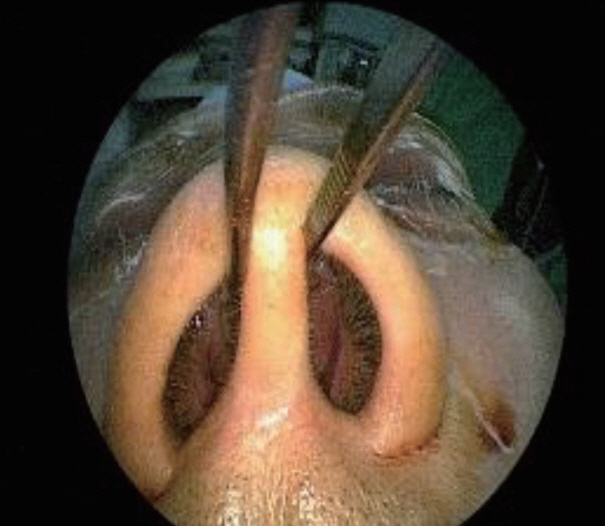
Fig. 1. Nasal endoscopic finding shows both nasal cavity obstructed by septal mass.
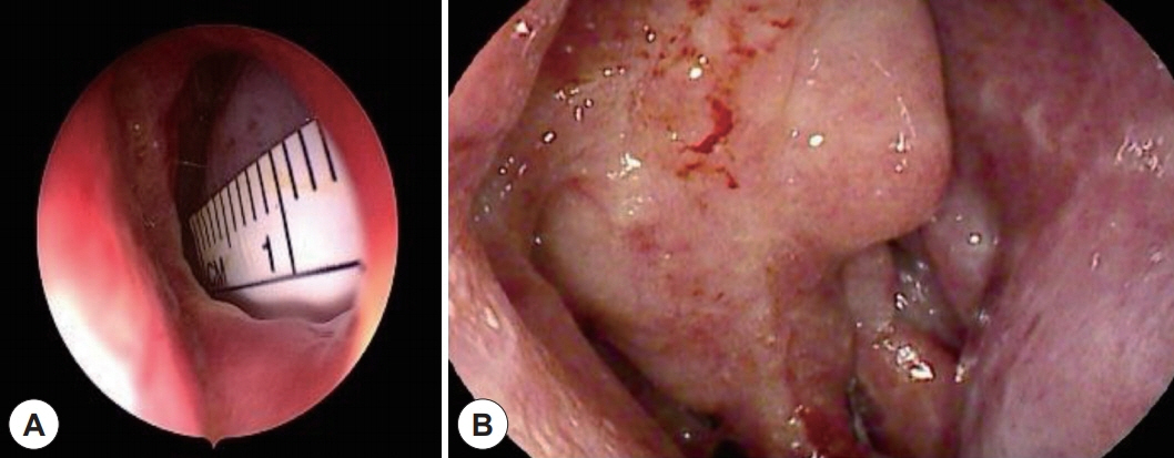
Fig. 2. Nasal endoscopic findings show a septal perforation (A) and a smooth-surfaced, pinkish-colored mass lesion on cephalic area of nasal septum (B).
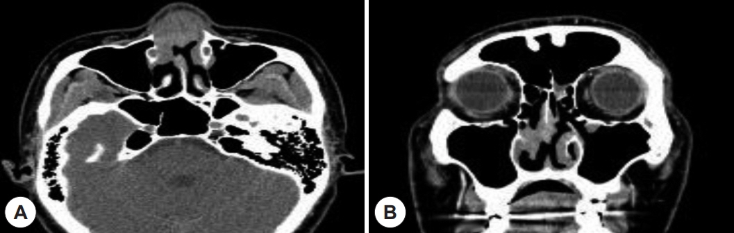
Fig. 3. PNS CT demonstrates a heterogeneous enhanced soft tissue mass at nasal dorsum and septum that extends to inferior turbinate. The axial view (A) and coronal view (B).
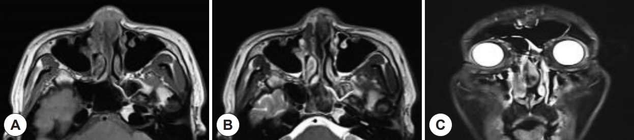
Fig. 4. Preoperative MRI finding. A: T1-weighted axial image demonstrates a heterogeneous enhanced mass with moderate signal intensity at nasal dorsum and septum. B: T2-weighted axial image demonstrates a heterogeneous enhanced mass with low signal intensity at nasal dorsum and septum. C: T1-weighted enhanced coronal image shows relatively well-marginated mass with high intensity signal at the septum.
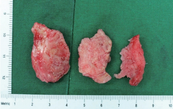
Fig. 5. Tumor is presented as a firm, well-circumscribed nodular mass with a various cut surface.

Fig. 6. Tumor is full of spindle cell arranged in fascicular pattern. Proliferation of fibrocollagenous tissue mixed with inflammatory cells including eosinophils, neutrophils and lymphocytes (H & E, ×100)(A). Histologic finding shows low cellurarity, lack of cellular atypia and dense inflammatory infiltrates (H & E, ×400)(B).
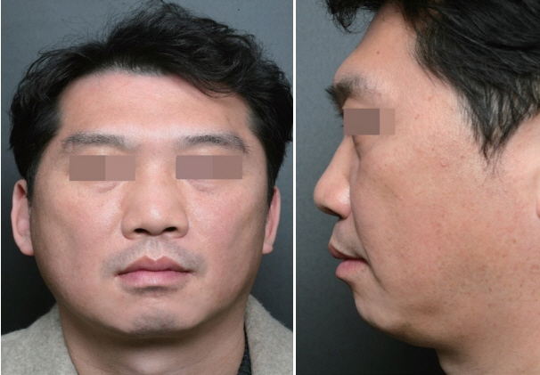
Fig. 7. The frontal and lateral views of patient shows no prominent saddle nose deformity after the operation.
Reference
-
References
1. Boudhas A, Allaoui M, El Asri F, Rharrassi I, El Ochi MR, Tbouda M, et al. Inflammatory myofibroblastic tumor of the lacrimal gland: case report of an exceptional location. BMC Clin Pathol. 2017; 17:12.2. Donner LR, Trompler RA, White RRt. Progression of inflammatory myofibroblastic tumor (inflammatory pseudotumor) of soft tissue into sarcoma after several recurrences. Hum Pathol. 1996; 27(10):1095–8.3. Som PM, Brandwein MS, Maldjian C, Reino AJ, Lawson W. Inflammatory pseudotumor of the maxillary sinus: CT and MR findings in six cases. AJR Am J Roentgenol. 1994; 163(3):689–92.4. He CY, Dong GH, Yang DM, Liu HG. Inflammatory myofibroblastic tumors of the nasal cavity and paranasal sinus: a clinicopathologic study of 25 cases and review of the literature. Eur Arch Otorhinolaryngol. 2015; 272(4):789–97.5. Lawrence B, Perez-Atayde A, Hibbard MK, Rubin BP, Dal Cin P, Pinkus JL, et al. TPM3-ALK and TPM4-ALK oncogenes in inflammatory myofibroblastic tumors. Am J Pathol. 2000; 157(2):377–84.6. Gale N, Zidar N, Podboj J, Volavsek M, Luzar B. Inflammatory myofibroblastic tumour of paranasal sinuses with fatal outcome: reactive lesion or tumour? J Clin Pathol. 2003; 56(9):715–7.7. Soysal V, Yigitbasi OG, Kontas O, Kahya HA, Guney E. Inflammatory myofibroblastic tumor of the nasal cavity: a case report and review of the literature. Int J Pediatr Otorhinolaryngol. 2001; 61(2):161–5.8. McDermott M. Inflammatory myofibroblastic tumour. Semin Diagn Pathol. 2016.9. Newlin HE, Werning JW, Mendenhall WM. Plasma cell granuloma of the maxillary sinus: a case report and literature review. Head Neck. 2005; 27(8):722–8.10. Coffin CM, Hornick JL, Fletcher CD. Inflammatory myofibroblastic tumor: comparison of clinicopathologic, histologic, and immunohistochemical features including ALK expression in atypical and aggressive cases. Am J Surg Pathol. 2007; 31(4):509–20.11. Coffin CM, Watterson J, Priest JR, Dehner LP. Extrapulmonary inflammatory myofibroblastic tumor (inflammatory pseudotumor). A clinicopathologic and immunohistochemical study of 84 cases. Am J Surg Pathol. 1995; 19(8):859–72.12. Karnak I, Senocak ME, Ciftci AO, Caglar M, Bingol-Kologlu M, Tanyel FC, et al. Inflammatory myofibroblastic tumor in children: diagnosis and treatment. J Pediatr Surg. 2001; 36(6):908–12.13. Okumura Y, Nomura K, Oshima T, Kasajima A, Suzuki T, Ishida E, et al. Inflammatory myofibroblastic tumor of the nasal septum. Case Rep Otolaryngol. 2013; 2013:670105.14. Polito E, Pichierri P, Loffredo A, Moramarco A, Occhini R. Inflammatory myofibroblastic tumor of the orbit. Ophthalmologica. 2007; 221(5):353–5.
- Full Text Links
-
- Actions
-
Cited
- CITED
-
- Close
- Share
-
- Similar articles
-
- A Case of Inflammatory Myofibroblastic Tumor of the Maxillary Sinus
- Inflammatory Myofibroblastic Tumor of the Nasal Cavity
- A Case of Inflammatory Myofibroblastic Tumor of Nasal Cavity in an 8 Year-Old Girl
- Glomus Tumor of the Nasal Septum : A Case Report
- The Effect of Nasal Septal Deviation and Septoplasty on Dacryocystorhinostomy Progression
