Restor Dent Endod.
2014 Feb;39(1):32-38.
Antibacterial effect of self-etching adhesive systems on Streptococcus mutans
- Affiliations
-
- 1Department of Conservative Dentistry, Dankook University College of Dentistry and Institute of Dental Science, Cheonan, Korea. donyshin@dankook.ac.kr
Abstract
OBJECTIVES
In this study, we evaluated the antibacterial activity of self-etching adhesive systems against Streptococcus mutans using the agar diffusion method.
MATERIALS AND METHODS
Three 2-step systems, Clearfil SE Bond (SE, Kuraray), Contax (CT, DMG), and Unifil Bond (UnB, GC), and three 1-step systems, Easy Bond (EB, 3M ESPE), U-Bond (UB, Vericom), and All Bond SE (AB, BISCO) were used. 0.12% chlorhexidine (CHX, Bukwang) and 37% phosphoric acid gel (PA, Vericom) were used as positive controls.
RESULTS
The antibacterial activity of CHX and PA was stronger than that of the other groups, except SE. After light activation, the inhibition zone was reduced in the case of all 2-step systems except CT. However, all 1-step systems did not exhibit any inhibition zone upon the light activation.
CONCLUSIONS
SE may be better than CT or UnB among the 2-step systems with respect to antibacterial activity, however, 1-step systems do not exhibit any antibacterial activity after light curing.
MeSH Terms
Figure
-
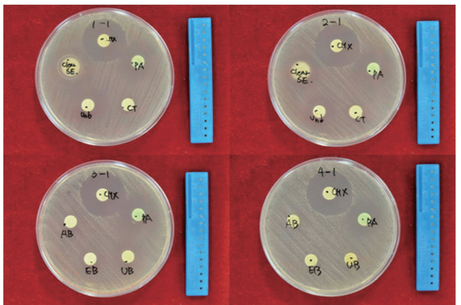
Figure 1 Inhibition zone of self-etching systems. CHX, Chlorhexidine; PA, Phosphoric acid; SE, Clearfil SE Bond; UnB, Unifil Bond; CT, Contax; AB, All Bond SE; EB, Easy Bond; UB, U-Bond. Plate 1-1, 3-1, No light activation; Plate 2-1, 4-1, Light activation.
Reference
-
1. Palotie U, Vehkalahti MM. Reasons for replacement of restorations: dentists' perceptions. Acta Odontol Scand. 2012; 70:485–490.
Article2. Hahn P, Weyen G, Fischer P, Plogmann S, Hannig M. Marginal and internal adaptation of composite restorations to dentin in vivo and in vitro. Am J Dent. 2008; 21:356–360.3. Lee MA, Seo DK, Son HH, Cho BH. Influence of rebonding procedures on microleakage of composite resin restorations. J Korean Acad Conserv Dent. 2010; 35:164–172.
Article4. Kidd EA, Joyston-Bechal S, Beighton D. Microbiological validation of assessments of caries activity during cavity preparation. Caries Res. 1993; 27:402–408.
Article5. Yip HK, Stevenson AG, Beeley JA. The specificity of caries detector dyes in cavity preparation. Br Dent J. 1994; 176:417–421.
Article6. Brännström M, Nordenvall KJ. Bacterial penetration, pulpal reaction and the inner surface of Concise enamel bond. Composite fillings in etched and unetched cavities. J Dent Res. 1978; 57:3–10.7. Ricketts DN, Kidd EA, Beighton D. Operative and microbiological validation of visual, radiographic and electronic diagnosis of occlusal caries in non-cavitated teeth judged to be in need of operative care. Br Dent J. 1995; 179:214–220.
Article8. Kim JS, Shin DH. Inhibitory effect on Streptococcus mutans and mechanical properties of the chitosan containing composite resin. Restor Dent Endod. 2013; 38:36–42.
Article9. Imazato S, Kinomoto Y, Tarumi H, Torii M, Russell RR, McCabe JF. Incorporation of antibacterial monomer MDPB into dentin primer. J Dent Res. 1997; 76:768–772.
Article10. Settembrini L, Boylan R, Strassler H, Scherer W. A comparison of antimicrobial activity of etchants used for a total etch technique. Oper Dent. 1997; 22:84–88.11. Imazato S, Torii Y, Takatsuka T, Inoue K, Ebi N, Ebisu S. Bactericidal effect of dentin primer containing antibacterial monomer methacryloyloxydodecylpyridini um bromide (MDPB) against bacteria in human carious dentin. J Oral Rehabil. 2001; 28:314–319.
Article12. Imazato S, Kuramoto A, Takahashi Y, Ebisu S, Peters MC. In vitro antibacterial effects of the dentin primer of Clearfil Protect Bond. Dent Mater. 2006; 22:527–532.
Article13. Karanika-Kouma A, Dionysopoulos P, Koliniotou-Koubia E, Kolokotronis A. Antibacterial properties of dentin bonding systems, polyacid-modified composite resins and composite resins. J Oral Rehabil. 2001; 28:157–160.
Article14. Peris AR, Mitsui FH, Lobo MM, Bedran-russo AK, Marchi GM. Adhesive systems and secondary caries formation: assessment of dentin bond strength, caries lesions depth and fluoride release. Dent Mater. 2007; 23:308–316.
Article15. Kim S, Song M, Roh BD, Park SH, Park JW. Inhibition of Streptococcus mutans biofilm formation on composite resins containing ursolic acid. Restor Dent Endod. 2013; 38:65–72.
Article16. Walter R, Duarte WR, Pereira PN, Heymann HO, Swift EJ Jr, Arnold RR. In vitro inhibition of bacterial growth using different dental adhesive systems. Oper Dent. 2007; 32:388–393.
Article17. Imazato S. Antibacterial properties of resin composites and dentin bonding systems. Dent Mater. 2003; 19:449–457.
Article18. Ohmori K, Maeda N, Kohno A. Evaluation of antibacterial activity of three dentin primers using an in vitro tooth model. Oper Dent. 1999; 24:279–285.19. Gondim JO, Duque C, Hebling J, Giro EM. Influence of human dentine on the antibacterial activity of self-etching adhesive systems against cariogenic bacteria. J Dent. 2008; 36:241–248.
Article20. Tobias RS. Antibacterial properties of dental restorative materials: a review. Int Endod J. 1988; 21:155–160.
Article21. Abdulkader A, Duguid R, Saunders EM. The antimicrobial activity of endodontic sealers to anaerobic bacteria. Int Endod J. 1996; 29:280–283.
Article22. Siqueira JF Jr, Favieri A, Gahyva SM, Moraes SR, Lima KC, Lopes HP. Antimicrobial activity and flow rate of newer and established root canal sealers. J Endod. 2000; 26:274–277.
Article23. Loesche WJ. Role of Streptococcus mutans in human dental decay. Microbiol Rev. 1986; 50:353–380.24. Hamada S, Koga T, Ooshima T. Virulence factors of Streptococcus mutans and dental caries prevention. J Dent Res. 1984; 63:407–411.
Article25. Fitzgerald RJ, Keyes PH. Demonstration of the etiologic role of streptococci in experimental caries in the hamster. J Am Dent Assoc. 1960; 61:9–19.
Article26. Bender GR, Thibodeau EA, Marquis RE. Reduction of acidurance of streptococcal growth and glycolysis by fluoride and gramicidin. J Dent Res. 1985; 64:90–95.
Article27. Lemos JA, Burne RA. A model of efficiency: stress tolerance by Streptococcus mutans. Microbiology. 2008; 154:3247–3255.28. Banas JA. Virulence properties of Streptococcus mutans. Front Biosci. 2004; 9:1267–1277.29. Bapna MS, Mukherjee S, Murphy R. The antimicrobial effect of an iron-binding agent on Streptococcus mutans. J Oral Rehabil. 1992; 19:111–113.
Article30. Kudou Y, Obara K, Kawashima T, Kubota M, Abe S, Endo T, Komatsu M, Okuda R. Addition of antibacterial agents to MMA-TBB dentin bonding systems-influences on tensile bond strength and antibacterial effect. Dent Mater J. 2000; 19:65–74.
Article31. Feuerstein O, Matalon S, Slutzky H, Weiss EI. Anti-bacterial properties of self-etching dental adhesive systems. J Am Dent Assoc. 2007; 138:349–354.
Article32. Schmalz G, Ergücü Z, Hiller KA. Effect of dentin on the antibacterial activity of dentin bonding agents. J Endod. 2004; 30:352–358.
Article33. Imazato S, Kuramoto A, Kaneko T, Ebisu S, Russell RR. Comparison of antibacterial activity of simplified adhesive systems. Am J Dent. 2002; 15:356–360.34. Cadenaro M, Antoniolli F, Sauro S, Tay FR, Di Lenarda R, Prati C, Biasotto M, Contardo L, Breschi L. Degree of conversion and permeability of dental adhesives. Eur J Oral Sci. 2005; 113:525–530.
Article35. Finger WJ, Lee KS, Podszun W. Monomers with low oxygen inhibition as enamel/dentin adhesives. Dent Mater. 1996; 12:256–261.
Article
- Full Text Links
-
- Actions
-
Cited
- CITED
-
- Close
- Share
-
- Similar articles
-
- Inhibitory effect of mastic oil on Streptococcus mutans growth
- Effects of Diospyros kaki peel, Momordica charantia, and Canavalia gladiata extracts on the cariogenic traits of Streptococcus mutans
- Identification and partial purification of antibacterial compounds against Streptococcus mutans from Galla Rhois
- Inhibitory effect on Streptococcus mutans and mechanical properties of the chitosan containing composite resin
- Galla chinensis extracts and calcium induce remineralization and antibacterial effects of enamel in a Streptococcus mutans biofilm model
