Pediatr Gastroenterol Hepatol Nutr.
2015 Mar;18(1):55-59. 10.5223/pghn.2015.18.1.55.
Achalasia Previously Diagnosed as Gastroesophageal Reflux Disease by Relying on Esophageal Impedance-pH Monitoring: Use of High-Resolution Esophageal Manometry in Children
- Affiliations
-
- 1Department of Pediatrics, Korea University College of Medicine, Seoul, Korea. shimjo@korea.ac.kr
- KMID: 2315536
- DOI: http://doi.org/10.5223/pghn.2015.18.1.55
Abstract
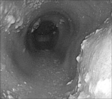
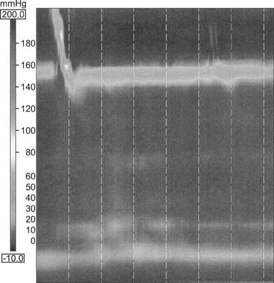
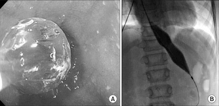
- Gastroesophageal reflux disorder (GERD) is the most common esophageal disorder in children. Achalasia occurs less commonly but has similar symptoms to GERD. A nine-year old boy presented with vomiting, heartburn, and nocturnal cough. The esophageal impedance-pH monitor revealed nonacidic GERD (all-refluxate clearance percent time of 20.9%). His symptoms persisted despite medical treatment for GERD, and he was lost to follow up. Four years later, he presented with heartburn, solid-food dysphagia, daily post-prandial vomiting, and failure to thrive. Endoscopy showed a severely dilated esophagus with candidiasis. High-resolution manometry was performed, and he was diagnosed with classic achalasia (also known as type I). His symptoms resolved after two pneumatic dilatation procedures, and his weight and height began to catch up to his peers. Clinicians might consider using high-resolution manometry in children with atypical GERD even after evaluation with an impedance-pH monitor.
Keyword
MeSH Terms
Figure
Reference
-
1. Vakil N, van Zanten SV, Kahrilas P, Dent J, Jones R. Global Consensus Group. The Montreal definition and classification of gastroesophageal reflux disease: a global evidence-based consensus. Am J Gastroenterol. 2006; 101:1900–1920.
Article2. Lightdale JR, Gremse DA. Section on Gastroenterology, Hepatology, and Nutrition. Gastroesophageal reflux: management guidance for the pediatrician. Pediatrics. 2013; 131:e1684–e1695.
Article3. Rosen R, Nurko S. The importance of multichannel intraluminal impedance in the evaluation of children with persistent respiratory symptoms. Am J Gastroenterol. 2004; 99:2452–2458.
Article4. Shoenut JP, Micflikier AB, Yaffe CS, Den Boer B, Teskey JM. Reflux in untreated achalasia patients. J Clin Gastroenterol. 1995; 20:6–11.
Article5. Smart HL, Mayberry JF, Atkinson M. Achalasia following gastro-oesophageal reflux. J R Soc Med. 1986; 79:71–73.
Article6. Franklin AL, Petrosyan M, Kane TD. Childhood achalasia: A comprehensive review of disease, diagnosis and therapeutic management. World J Gastrointest Endosc. 2014; 6:105–111.
Article7. Kahrilas PJ, Sifrim D. High-resolution manometry and impedance-pH/manometry: valuable tools in clinical and investigational esophagology. Gastroenterology. 2008; 135:756–769.
Article8. Dellon ES. Eosinophilic esophagitis. Gastroenterol Clin North Am. 2013; 42:133–153.
Article9. Roskies M, Zielinski D, Levesque D, Daniel SJ. Atypical presentations of achalasia in the pediatric population. J Otolaryngol Head Neck Surg. 2012; 41:E44–E46.10. Kahrilas PJ, Boeckxstaens G. The spectrum of achalasia: lessons from studies of pathophysiology and high-resolution manometry. Gastroenterology. 2013; 145:954–965.
Article11. Ganatra JV, Bostwick HE, Medow MS, Beneck D, Berezin S. Candida esophagitis in a child with achalasia. J Pediatr Gastroenterol Nutr. 1996; 22:330–333.
Article12. Cools-Lartigue J, Chang SY, Mckendy K, Mayrand S, Marcus V, Fried GM, et al. Pattern of esophageal eosinophilic infiltration in patients with achalasia and response to Heller myotomy and Dor fundoplication. Dis Esophagus. 2013; 26:766–775.
Article13. Dellon ES. Diagnostics of eosinophilic esophagitis: clinical, endoscopic, and histologic pitfalls. Dig Dis. 2014; 32:48–53.
Article14. Loots CM, Benninga MA, Omari TI. Gastroesophageal reflux in pediatrics; (patho)physiology and new insights in diagnostics and treatment. Minerva Pediatr. 2012; 64:101–119.15. Bredenoord AJ, Fox M, Kahrilas PJ, Pandolfino JE, Schwizer W, Smout AJ. International High Resolution Manometry Working Group. Chicago classification criteria of esophageal motility disorders defined in high resolution esophageal pressure topography. Neurogastroenterol Motil. 2012; 24:Suppl 1. 57–65.
Article


