Assessment of the anterior loop of the mandibular canal: A study using cone-beam computed tomography
- Affiliations
-
- 1Department of Oral Diagnosis, Division of Oral Radiology, Piracicaba Dental School, University of Campinas (UNICAMP), São Paulo, Brazil.
- 2Department of Clinical and Preventive Dentistry, School of Dentistry, Federal University of Pernambuco (UFPE), Recife, Pernambuco, Brazil. flavia_radio@hotmail.com
- 3Instituto de Medicina Integral Professor Fernando Figueira - IMIP, Recife, Pernambuco, Brazil.
- 4Division of Oral Radiology, Recife Dental School (FOR), Recife, Pernambuco, Brazil.
- KMID: 2308869
- DOI: http://doi.org/10.5624/isd.2016.46.2.69
Abstract
- PURPOSE
Sufficient area in the interforaminal region is required for dental implant placement, and the anterior loop of the mandibular canal is located within the limits of this area. The aim of this study was to evaluate the prevalence and extent of the anterior loop in a Brazilian sample population using cone-beam computed tomography (CBCT).
MATERIALS AND METHODS
CBCT images from 250 patients (500 hemimandibles) obtained for various clinical indications were randomly selected and evaluated to determine the presence and length of the anterior loop. The length of the anterior loop was then compared based on gender, age, and the side of the mandible. The data were analyzed using the Pearson chi-square test and linear regression analysis.
RESULTS
An anterior loop was identified in 41.6% of the cases, and its length ranged from 0.25 mm to 4.00 mm (mean, 1.1±0.8 mm). The loop had a greater mean length and was significantly more prevalent in males (p=0.014). No significant differences were found between the right and left sides regarding length (p=0.696) or prevalence (p=0.650).
CONCLUSION
In this study, a high prevalence of the anterior loop of the mandibular canal was found, and although its length varied greatly, in most cases it was less than 1 mm long. Although this is a prevalent anatomical variation, safety limits for the placement of implants in this region cannot be established before an accurate evaluation using imaging techniques in order to identify and preserve the neurovascular bundles.
MeSH Terms
Figure
-
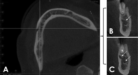
Fig. 1 Assessment of the anterior loop. A. Axial reconstruction rotated towards the side being examined, with the mandibular canal positioned parallel to the sagittal plane (vertical line). B and C. The coronal reconstructions obtained immediately after the most mesial point of the mental foramen show a single (arrow) and double (arrows) hypodense image, respectively.
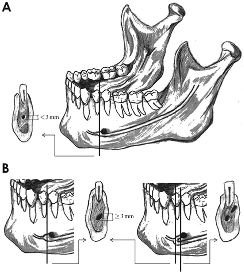
Fig. 2 The course of the mandibular canal, with the absence or presence of an anterior loop. A. The incisive canal and its corresponding image in the coronal reconstruction, with a diameter that must be less than 3 mm. B. The anterior loop and its corresponding image in the coronal reconstruction, which, depending on the place it is obtained, can be represented by a round hypodense area with a diameter of at least 3 mm or by two round hypodense areas.
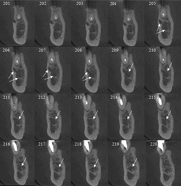
Fig. 3 In coronal reconstructions, the anterior loop can be seen as two round hypodense areas in images 205-208 and as a single round hypodense area (with a diameter of at least 3 mm) in images 209-219. Measurement: 15×0.25=3.75 mm.
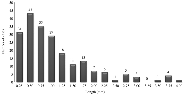
Fig. 4 The number of cases of anterior loop of the mandibular canal and their respective lengths.
Reference
-
1. Apostolakis D, Brown JE. The anterior loop of the inferior alveolar nerve: prevalence, measurement of its length and a recommendation for interforaminal implant installation based on cone beam CT imaging. Clin Oral Implants Res. 2012; 23:1022–1030.
Article2. Rosa MB, Sotto-Maior BS, Machado Vde C, Francischone CE. Retrospective study of the anterior loop of the inferior alveolar nerve and the incisive canal using cone beam computed tomography. Int J Oral Maxillofac Implants. 2013; 28:388–392.
Article3. Filo K, Schneider T, Locher MC, Kruse AL, Lübbers H. The inferior alveolar nerve's loop at the mental foramen and its implications for surgery. J Am Dent Assoc. 2014; 145:260–269.
Article4. Greenstein G, Tarnow D. The mental foramen and nerve: clinical and anatomical factors related to dental implant placement: a literature review. J Periodontol. 2006; 77:1933–1943.
Article5. Kaya Y, Sencimen M, Sahin S, Okcu KM, Dogan N, Bahcecitapar M. Retrospective radiographic evaluation of the anterior loop of the mental nerve: comparison between panoramic radiography and spiral computerized tomography. Int J Oral Maxillofac Implants. 2008; 23:919–925.6. de Oliveira-Santos C, Souza PH, de Azambuja Berti-Couto S, Stinkens L, Moyaert K, Rubira-Bullen IR, et al. Assessment of variations of the mandibular canal through cone beam computed tomography. Clin Oral Investig. 2012; 16:387–393.
Article7. Parnia F, Moslehifard E, Hafezeqoran A, Mahboub F, Mojaver-Kahnamoui H. Characteristics of anatomical landmarks in the mandibular interforaminal region: a cone-beam computed tomography study. Med Oral Patol Oral Cir Bucal. 2012; 17:e420–e425.
Article8. Uchida Y, Yamashita Y, Goto M, Hanihara T. Measurement of anterior loop length for the mandibular canal and diameter of the mandibular incisive canal to avoid nerve damage when installing endosseous implants in the interforaminal region. J Oral Maxillofac Surg. 2007; 65:1772–1779.
Article9. Uchida Y, Noguchi N, Goto M, Yamashita Y, Hanihara T, Takamori H, et al. Measurement of anterior loop length for the mandibular canal and diameter of the mandibular incisive canal to avoid nerve damage when installing endosseous implants in the interforaminal region: a second attempt introducing cone beam computed tomography. J Oral Maxillofac Surg. 2009; 67:744–750.
Article10. Neiva RF, Gapski R, Wang HL. Morphometric analysis of implant-related anatomy in Caucasian skulls. J Periodontol. 2004; 75:1061–1067.
Article11. Kuzmanovic DV, Payne AG, Kieser JA, Dias GJ. Anterior loop of the mental nerve: a morphological and radiographic study. Clin Oral Implants Res. 2003; 14:464–471.
Article12. Li X, Jin ZK, Zhao H, Yang K, Duan JM, Wang WJ. The prevalence, length and position of the anterior loop of the inferior alveolar nerve in Chinese, assessed by spiral computed tomography. Surg Radiol Anat. 2013; 35:823–830.
Article13. Liang X, Jacobs R, Hassan B, Li L, Pauwels R, Corpas L, et al. A comparative evaluation of cone beam computed tomography (CBCT) and multi-slice CT (MSCT) Part I. On subjective image quality. Eur J Radiol. 2010; 75:265–269.14. Suomalainen A, Kiljunen T, Kåser Y, Peltola J, Kortesniemi M. Dosimetry and image quality of four dental cone beam computed tomography scanners compared with multislice computed tomography scanners. Dentomaxillofac Radiol. 2009; 38:367–378.
Article15. Watanabe H, Mohammad Abdul M, Kurabayashi T, Aoki H. Mandible size and morphology determined with CT on a premise of dental implant operation. Surg Radiol Anat. 2010; 32:343–349.
Article16. Ngeow WC, Dionysius DD, Ishak H, Nambiar P. A radiographic study on the visualization of the anterior loop in dentate subjects of different age groups. J Oral Sci. 2009; 51:231–237.
Article
- Full Text Links
-
- Actions
-
Cited
- CITED
-
- Close
- Share
-
- Similar articles
-
- Observation of the anterior loop and mental foramen of the mandibular canal using cone beam computed tomography
- Assessment of the relationship between the mandibular third molar and the mandibular canal using panoramic radiograph and cone beam computed tomography
- Panoramic radiographs underestimate extensions of the anterior loop and mandibular incisive canal
- Correlation of panoramic radiographs and cone beam computed tomography in the assessment of a superimposed relationship between the mandibular canal and impacted third molars
- Observation of mandibular second molar roots and root canal morphology using dental cone-beam computed tomography
