Panoramic radiographs underestimate extensions of the anterior loop and mandibular incisive canal
- Affiliations
-
- 1Department of Oral Diagnosis, Division of Oral Radiology, Piracicaba Dental School, University of Campinas, São Paulo, Brazil. acarolinerb@hotmail.com
- 2Department of Stomatology, Public Oral Health and Forensic Dentistry, School of Dentistry of Ribeirão Preto, University of São Paulo, São Paulo, Brazil.
- KMID: 2408247
- DOI: http://doi.org/10.5624/isd.2016.46.3.159
Abstract
- PURPOSE
The purpose of this study was to detect the anterior loop of the mental nerve and the mandibular incisive canal in panoramic radiographs (PAN) and cone-beam computed tomography (CBCT) images, as well as to determine the anterior/mesial extension of these structures in panoramic and cross-sectional reconstructions using PAN and CBCT images.
MATERIALS AND METHODS
Images (both PAN and CBCT) from 90 patients were evaluated by 2 independent observers. Detection of the anterior loop and the incisive canal were compared between PAN and CBCT. The anterior/mesial extension of these structures was compared between PAN and both cross-sectional and panoramic CBCT reconstructions.
RESULTS
In CBCT, the anterior loop and the incisive canal were observed in 7.7% and 24.4% of the hemimandibles, respectively. In PAN, the anterior loop and the incisive canal were detected in 15% and 5.5% of cases, respectively. PAN presented more difficulties in the visualization of structures. The anterior/mesial extensions ranged from 0.0 mm to 19.0 mm on CBCT. PAN underestimated the measurements by approximately 2.0 mm.
CONCLUSION
CBCT appears to be a more reliable imaging modality than PAN for preoperative workups of the anterior mandible. Individual variations in the anterior/mesial extensions of the anterior loop of the mental nerve and the mandibular incisive canal mean that is not prudent to rely on a general safe zone for implant placement or bone surgery in the interforaminal region.
Figure
-
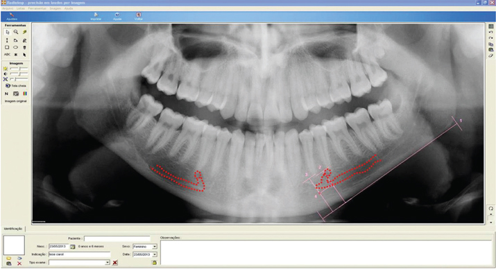
Fig. 1 A panoramic radiograph shows the anterior loop and incisive canal (right side) and the anterior loop (left side). 1. The lower mandibular cortex as the plane of reference. 2. The line perpendicular to the line passing through the mesial border of the mental foramen. 3. The line perpendicular to the line passing through the most mesial point of the anterior loop of the mental nerve with the mandibular incisive canal. 4. The distance between lines 2 and 3, corresponding to the mesial length of the extent of the anterior loop or incisive canal.
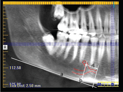
Fig. 2 Visualization of the anterior loop and incisive canal (red dotted line) on the panoramic reconstructions. a: The lower mandibular cortex as the plane of reference. b: The line perpendicular to the line passing through the mesial border of the mental foramen. c: The line perpendicular to the line passing through the most mesial point of the anterior loop of the mental nerve and the mandibular incisive canal. d: The distance between lines b and c, corresponding to the mesial length of the extent of the anterior loop or incisive canal.
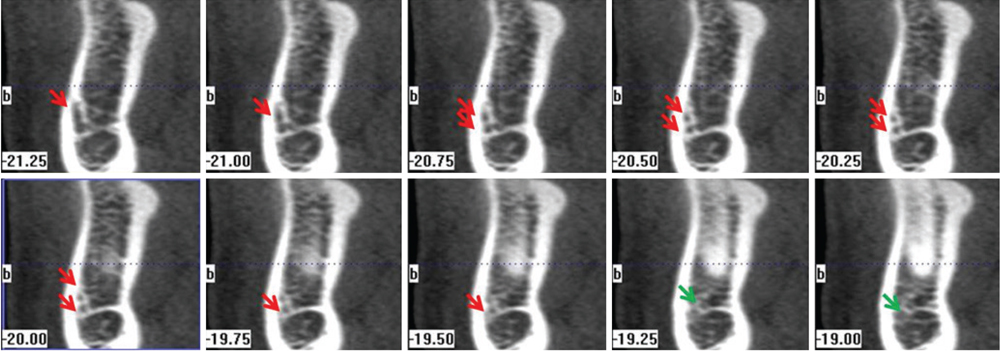
Fig. 3 Cross-sectional images (perpendicular to the occlusal plane) demonstrate the presence of the anterior loop (red arrows) and the incisive canal (green arrows).
Reference
-
1. Liang X, Lambrichts I, Corpas L, Politis C, Vrielinck L, Ma GW, et al. Neurovascular disturbance associated with implant placement in the anterior mandibular and its surgical implications: literature review including report of a case. Chin J Dent Res. 2008; 11:56–64.2. Mardinger O, Chaushu G, Arensburg B, Taicher S, Kaffe I. Anterior loop of the mental canal: an anatomical-radiologic study. Implant Dent. 2000; 9:120–125.3. Kuzmanovic DV, Payne AG, Kieser JA, Dias GJ. Anterior loop of the mental nerve: a morphological and radiographic study. Clin Oral Implants Res. 2003; 14:464–471.
Article4. Hu KS, Yun HS, Hur MS, Kwon HJ, Abe S, Kim HJ. Branching patterns and intraosseous course of the mental nerve. J Oral Maxillofac Surg. 2007; 65:2288–2294.
Article5. Uchida Y, Yamashita Y, Goto M, Hanihara T. Measurement of anterior loop length for the mandibular canal and diameter of the mandibular incisive canal to avoid nerve damage when installing endosseous implants in the interforaminal region. J Oral Maxillofac Surg. 2007; 65:1772–1779.
Article6. Uchida Y, Noguchi N, Goto M, Yamashita Y, Hanihara T, Takamori H, et al. Measurement of anterior loop length for the mandibular canal and diameter of the mandibular incisive canal to avoid nerve damage when installing endosseous implants in the interforaminal region: a second attempt introducing cone beam computed tomography. J Oral Maxillofac Surg. 2009; 67:744–750.
Article7. Ngeow WC, Dionysius DD, Ishak H, Nambiar P. A radiographic study on the visualization of the anterior loop in dentate subjects of different age groups. J Oral Sci. 2009; 51:231–237.
Article8. Benninger B, Miller D, Maharathi A, Carter W. Dental implant placement investigation: is the anterior loop of the mental nerve clinically relevant? J Oral Maxillofac Surg. 2011; 69:182–185.
Article9. de Oliveira-Santos C, Souza PH, de Azambuja Berti-Couto S, Stinkens L, Moyaert K, Rubira-Bullen IR, et al. Assessment of variations of the mandibular canal through cone beam computed tomography. Clin Oral Investig. 2012; 16:387–393.
Article10. Mardinger O, Chaushu G, Arensburg B, Taicher S, Kaffe I. Anatomic and radiologic course of the mandibular incisive canal. Surg Radiol Anat. 2000; 22:157–161.
Article11. Jacobs R, Mraiwa N, Van Steenberghe D, Sanderink G, Quirynen M. Appearance of the mandibular incisive canal on panoramic radiographs. Surg Radiol Anat. 2004; 26:329–333.
Article12. Mraiwa N, Jacobs R, Moerman P, Lambrichts I, van Steenberghe D, Quirynen M. Presence and course of the incisive canal in the human mandibular interforaminal region: two-dimensional imaging versus anatomical observations. Surg Radiol Anat. 2003; 25:416–423.
Article13. Jalili MR, Esmaeelinejad M, Bayat M, Aghdasi MM. Appearance of anatomical structures of mandible on panoramic radiographs in Iranian population. Acta Odontol Scand. 2012; 70:384–389.
Article14. Pires CA, Bissada NF, Becker JJ, Kanawati A, Landers MA. Mandibular incisive canal: cone beam computed tomography. Clin Implant Dent Relat Res. 2012; 14:67–73.
Article15. Jacobs R, Mraiwa N, vanSteenberghe D, Gijbels F, Quirynen M. Appearance, location, course, and morphology of the mandibular incisive canal: an assessment on spiral CT scan. Dentomaxillofac Radiol. 2002; 31:322–327.
Article16. Makris N, Stamatakis H, Syriopoulos K, Tsiklakis K, van der Stelt PF. Evaluation of the visibility and the course of the mandibular incisive canal and the lingual foramen using conebeam computed tomography. Clin Oral Implants Res. 2010; 21:766–771.
Article17. Sokhn S, Nasseh I, Noujeim M. Using cone beam computed tomography to determine safe regions for implant placement. Gen Dent. 2011; 59:e72–e77.18. Parnia F, Moslehifard E, Hafezeqoran A, Mahboub F, Mojaver-Kahnamoui H. Characteristics of anatomical landmarks in the mandibular interforaminal region: a cone-beam computed tomography study. Med Oral Patol Oral Cir Bucal. 2012; 17:e420–e425.
Article19. Kajan ZD, Salari A. Presence and course of the mandibular incisive canal and presence of the anterior loop in cone beam computed tomography images of an Iranian population. Oral Radiol. 2012; 28:55–61.
Article20. Apostolakis D, Brown JE. The dimensions of the mandibular incisive canal and its spatial relationship to various anatomical landmarks of the mandible: a study using cone beam computed tomography. Int J Oral Maxillofac Implants. 2013; 28:117–124.
Article21. Al-Ani O, Nambiar P, Ha KO, Ngeow WC. Safe zone for bone harvesting from the interforaminal region of the mandible. Clin Oral Implants Res. 2013; 24:Suppl A100. 115–121.
Article22. Kaya Y, Sencimen M, Sahin S, Okcu KM, Dogan N, Bahcecitapar M. Retrospective radiographic evaluation of the anterior loop of the mental nerve: comparison between panoramic radiography and spiral computerized tomography. Int J Oral Maxillofac Implants. 2008; 23:919–925.23. Kilic C, Kamburoğlu K, Ozen T, Balcioglu HA, Kurt B, Kutoglu T, et al. The position of the mandibular canal and histologic feature of the inferior alveolar nerve. Clin Anat. 2010; 23:34–42.
Article24. Landis JR, Koch GG. The measurement of observer agreement for categorical data. Biometrics. 1977; 33:159–174.
Article25. Liang X, Jacobs R, Corpas LS, Semal P, Lambrichts I. Chronologic and geographic variability of neurovascular structures in the human mandible. Forensic Sci Int. 2009; 190:24–32.
Article26. Couto-Filho CE, Moraes PH, Alonso MB, Haiter-Neto F, Olate S, Albergaria-Barbora JR. Accuracy in the diagnosis of the mental nerve loop. A comarative study between panoramic radiography and cone beam computed tomography. Int J Morphol. 2015; 33:327–332.27. Kieser J, Kuzmanovic D, Payne A, Dennison J, Herbison P. Patterns of emergence of the human mental nerve. Arch Oral Biol. 2002; 47:743–747.
Article28. Rosenquist B. Is there an anterior loop of the inferior alveolar nerve? Int J Periodontics Restorative Dent. 1996; 16:40–45.29. Rosa MB, Sotto-Maior BS, Machado Vde C, Francischone CE. Retrospective study of the anterior loop of the inferior alveolar nerve and the incisive canal using cone beam computed tomography. Int J Oral Maxillofac Implants. 2013; 28:388–392.
Article30. Apostolakis D, Brown JE. The anterior loop of the inferior alveolar nerve: prevalence, measurement of its length and a recommendation for interforaminal implant installation based on cone beam CT imaging. Clin Oral Implants Res. 2012; 23:1022–1030.
Article
- Full Text Links
-
- Actions
-
Cited
- CITED
-
- Close
- Share
-
- Similar articles
-
- Erratum to: Panoramic radiographs underestimate extensions of the anterior loop and mandibular incisive canal
- Morphological assessment of the anterior loop of the mandibular canal in Koreans
- Radiographic evaluation of the course and visibility of the mandibular canal
- Assessment of the relationship between the mandibular third molar and the mandibular canal using panoramic radiograph and cone beam computed tomography
- Correlation of panoramic radiographs and cone beam computed tomography in the assessment of a superimposed relationship between the mandibular canal and impacted third molars
