Abnormal Vestibular Evoked Myogenic Potentials in Medial Medullary Infarction
- Affiliations
-
- 1Department of Neurology, Seoul National University Bundang Hospital, Seoul National University College of Medicine, Seongnam, Korea. jisookim@snu.ac.kr
- 2Department of Otolaryngology and Head & Neck Surgery, Seoul National University Bundang Hospital, Seoul National University College of Medicine, Seongnam, Korea.
- 3Department of Neurology, Wonkwang University School of Medicine, Iksan, Korea.
- KMID: 2287663
- DOI: http://doi.org/10.3988/jcn.2009.5.2.101
Abstract
-
BACKGROUND: The medial vestibulospinal tract (MVST), which descends in the medial longitudinal fasciculus (MLF), may mediate the vestibular evoked myogenic potentials (VEMPs) in the contracting sternocleidomastoid muscle. We report herein abnormal VEMPs in a patient with medial medullary infarction (MMI) that appeared to involve the MLF.
CASE REPORT
A patient with infarction involving the right medial medulla showed decreased p13-n23 amplitude and increased p13/n23 latencies of the VEMPs on the right side. These abnormal VEMPs recorded in an MMI patient support the theory that VEMPs are mediated by the MVST contained within the MLF.
CONCLUSIONS
VEMPs may represent a valuable tool for investigating vestibular dysfunction originating from the saccule, even in patients with central vestibulopathies, which is not readily defined by conventional vestibular function tests.
MeSH Terms
Figure
-
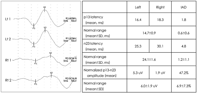
Fig. 1 Vestibular evoked myogenic potentials (VEMPs) recorded from the contracting ipsilateral sternocleidomastoid muscle exhibit decreased p13-n23 amplitude and increased p13/n23 latencies on the right side. The stimuli were short, alternating tone bursts (95 dB nHL, 108 dB SPL; 500 Hz; ramp=2 ms; plateau=3 ms) presented at 2.1 Hz monaurally. To compare the normalized p13-n23 amplitude of VEMP responses on the affected side with those on the intact side, the interaural difference ratio (IAD) of the normalized amplitude (IADamp, %) was calculated using [(Au-Aa)/(Aa+Au)×100], where Au is the normalized p13-n23 amplitude on the unaffected side and Aa is the amplitude on the affected side. Normative data were obtained from 52 healthy volunteers (23 women) aged between 22 and 76 years (36.7±2.4 years, mean±SD; median=32 years). Lt: left, Rt: right.
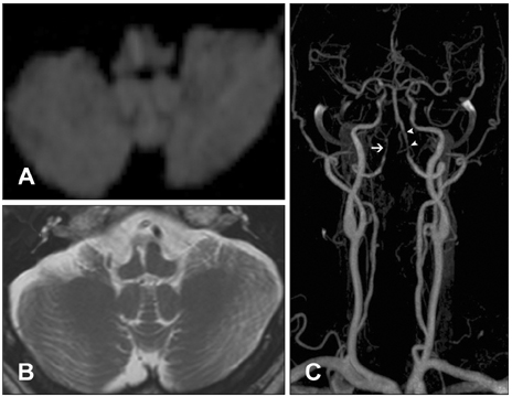
Fig. 2 Brain MRI of the patient with medial medullary infarction. A: Diffusion-weighted axial image disclosing acute infarction in the right medial medulla, which extends from the pyramid to the medullary tegmentum. B: Follow-up T2-weighted axial image also shows infarction in the corresponding area of the acute infarction shown on A. C: MR angiography revealing diffuse narrowing of the right distal vertebral artery (arrow) and focal narrowings in the left vertebral artery (arrowheads). MR: magnetic resonance.
Reference
-
1. Colebatch JG, Halmagyi GM. Vestibular evoked potentials in human neck muscles before and after unilateral vestibular deafferentation. Neurology. 1992. 42:1635–1636.
Article2. Colebatch JG, Halmagyi GM, Skuse NF. Myogenic potentials generated by a click-evoked vestibulocollic reflex. J Neurol Neurosurg Psychiatry. 1994. 57:190–197.
Article3. Murofushi T, Matsuzaki M, Wu CH. Short tone burst-evoked myogenic potentials on the sternocleidomastoid muscle: are these potentials also of vestibular origin? Arch Otolaryngol Head Neck Surg. 1999. 125:660–664.
Article4. Welgampola MS, Colebatch JG. Characteristics and clinical applications of vestibular-evoked myogenic potentials. Neurology. 2005. 64:1682–1688.
Article5. Murofushi T, Curthoys IS, Topple AN, Colebatch JG, Halmagyi GM. Responses of guinea pig primary vestibular neurons to clicks. Exp Brain Res. 1995. 103:174–178.
Article6. Kim JS, Choi KD, Oh SY, Park SH, Han MK, Yoon BW, et al. Medial medullary infarction: abnormal ocular motor findings. Neurology. 2005. 65:1294–1298.
Article7. Chen CH, Young YH. Vestibular evoked myogenic potentials in brainstem stroke. Laryngoscope. 2003. 113:990–993.
Article8. Fernández C, Goldberg JM. Physiology of peripheral neurons innervating otolith organs of the squirrel monkey. I. Response to static tilts and to long-duration centrifugal force. J Neurophysiol. 1976. 39:970–984.
Article9. Newlands SD, Vrabec JT, Purcell IM, Stewart CM, Zimmerman BE, Perachio AA. Central projections of the saccular and utricular nerves in macaques. J Comp Neurol. 2003. 466:31–47.
Article10. Murofushi T, Halmagyi GM, Yavor RA, Colebatch JG. Absent vestibular evoked myogenic potentials in vestibular neurolabyrinthitis. An indicator of inferior vestibular nerve involvement? Arch Otolaryngol Head Neck Surg. 1996. 122:845–848.
Article11. Robertson DD, Ireland DJ. Vestibular evoked myogenic potentials. J Otolaryngol. 1995. 24:3–8.12. Wilson VJ, Boyle R, Fukushima K, Rose PK, Shinoda Y, Sugiuchi Y, et al. The vestibulocollic reflex. J Vestib Res. 1995. 5:147–170.
Article13. Pompeiano O, Brodal A. The origin of vestibulospinal fibres in the cat. An experimental-anatomical study with comments on the descending medial longitudinal fasciculus. Arch Ital Biol. 1957. 95:166–195.14. Akaike T. Neuronal organization of the vestibulospinal system in the cat. Brain Res. 1983. 259:217–227.
Article
- Full Text Links
-
- Actions
-
Cited
- CITED
-
- Close
- Share
-
- Similar articles
-
- The Principle and Methodology of Vestibular Evoked Myogenic Potential
- Vestibular-evoked myogenic potentials: principle and clinical findings
- Cervical Vestibular Evoked Myogenic Potential and Ocular Vestibular Evoked Myogenic Potential in Patients With Vestibular Neuritis and Acute Viral Labyrinthitis
- Clinical Applications of Vestibular Evoked Myogenic Potentials in Vestibular Disorders
- Ocular Vestibular Evoked Myogenic Potential in Vestibular Neuritis Patients: Comparative Study with Cervical Vestibular Evoked Myogenic Potential and Subjective Visual Vertical
