Exofocal Anterograde Transsynaptic Neuronal Death in the Globus Pallidus: Two Case Reports
- Affiliations
-
- 1Department of Neurology, Hallym University College of Medicine, Seoul, Korea. yangki2@unitel.co.kr
- KMID: 2287584
- DOI: http://doi.org/10.3988/jcn.2012.8.4.308
Abstract
- BACKGROUND
Exofocal neuronal death in the substantia nigra (SN) is a well-known form of anterograde transsynaptic cell death. Exofocal neuronal death could theoretically also occur in the globus pallidus (GP) after striatal injury.
CASE REPORT
Case 1. A 70-year-old woman visited the emergency room because of decreased mentality. On admission, blood-gas analysis indicated that her oxygen tension was 69.1 mm Hg. The caudate nucleus, putamen, and temporooccipital cortex on both sides of the brain exhibited high-intensity diffusion-weighted magnetic resonance imaging (MRI) signals. At 10 days after admission, new high-intensity signals had developed in the SN and GP on both sides. Case 2. A 48-year-old man visited the emergency room because of right-sided weakness. Lesions were noted in the left caudate nucleus and putamen. At 4 days after admission, newly developed high-intensity MRI signals were observed in the left SN and GP.
CONCLUSIONS
Exofocal neuronal death can occur in the GP as well as in the SN; these findings need to be clearly distinguished from those of recurrent ischemic injuries, such as recurrent stroke.
Keyword
MeSH Terms
Figure
-
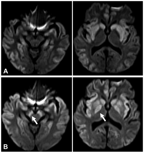
Fig. 1 Case 1. A: Diffusion-weighted magnetic resonance imaging scan of the brain on day 1, showing high-intensity signals in the caudate nucleus, putamen, and temporooccipital cortex on both sides of the brain. B: Follow-up diffusion-weighted magnetic resonance imaging scan performed 10 days after admission, showing newly developed high-intensity signals in the substantia nigra and globus pallidus (arrows).
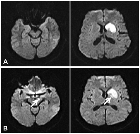
Fig. 2 Case 2. A: Diffusion-weighted brain magnetic resonance imaging scan performed on day 2 after admission, showing high-intensity signal lesions in the caudate nucleus and putamen. B: Newly developed high-intensity signals in the right substantia nigra and globus pallidus were observed on a diffusion-weighted magnetic resonance imaging image obtained 4 days after admission.
Reference
-
1. Zhao F, Kuroiwa T, Miyasaka N, Nagaoka T, Nakane M, Tamura A, et al. Ultrastructural and MRI study of the substantia nigra evolving exofocal post-ischemic neuronal death in the rat. Neuropathology. 2002. 22:91–105.
Article2. Zhao F, Kuroiwa T, Miyasaka N, Nagaoka T, Nakane M, Tamura A, et al. Characteristic changes in T(2)-value, apparent diffusion coefficient, and ultrastructure of substantia nigra evolving exofocal postischemic neuronal death in rats. Brain Res. 2001. 895:238–244.
Article3. Nakane M, Teraoka A, Asato R, Tamura A. Degeneration of the ipsilateral substantia nigra following cerebral infarction in the striatum. Stroke. 1992. 23:328–332.
Article4. Nambu A. Globus pallidus internal segment. Prog Brain Res. 2007. 160:135–150.
Article5. Kinoshita T, Sugihara S, Matsusue E, Fujii S, Ametani M, Ogawa T. Pallidoreticular damage in acute carbon monoxide poisoning: diffusion-weighted MR imaging findings. AJNR Am J Neuroradiol. 2005. 26:1845–1848.6. Gutierrez LG, Rovira A, Portela LA, Leite Cda C, Lucato LT. CT and MR in non-neonatal hypoxic-ischemic encephalopathy: radiological findings with pathophysiological correlations. Neuroradiology. 2010. 52:949–976.
Article7. Kita H, Tachibana Y, Nambu A, Chiken S. Balance of monosynaptic excitatory and disynaptic inhibitory responses of the globus pallidus induced after stimulation of the subthalamic nucleus in the monkey. J Neurosci. 2005. 25:8611–8619.
Article8. Tachibana Y, Kita H, Chiken S, Takada M, Nambu A. Motor cortical control of internal pallidal activity through glutamatergic and GAB-Aergic inputs in awake monkeys. Eur J Neurosci. 2008. 27:238–253.
Article9. Fève AP, Fénelon G, Wallays C, Rémy P, Guillard A. Axial motor disturbances after hypoxic lesions of the globus pallidus. Mov Disord. 1993. 8:321–326.
Article10. Gerfen CR. Molecular effects of dopamine on striatal-projection pathways. Trends Neurosci. 2000. 23:S64–S70.
Article
- Full Text Links
-
- Actions
-
Cited
- CITED
-
- Close
- Share
-
- Similar articles
-
- MR Imaging of Kernicterus: A Case Report
- Globus Pallidus Lesions Associated with High Mountain Climbing
- Globus Pallidus Interna Deep Brain Stimulation for Chorea-Acanthocytosis
- Deep Brain Stimulation of the Globus Pallidus in a 7-Year-Old Girl with DYT1 Generalized Dystonia
- Computed tomography of delayed encephalopathy of acute carbon monoxide poisoning: correlation with clinicalfindings
