Pharyngeal airway analysis of different craniofacial morphology using cone-beam computed tomography (CBCT)
- Affiliations
-
- 1Department of Orthodontics, School of Dentistry, Pusan National University, Busan, Korea. sbypark@pusan.ac.kr
- KMID: 2273208
- DOI: http://doi.org/10.4041/kjod.2009.39.3.136
Abstract
OBJECTIVE
CBCT has become popular for orthodontic diagnosis and treatment planning in recent times. The 3D pharyngeal airway space needs to be analysed using a 3D diagnostic tool. The aim of this study was to analyse the pharyngeal airway of different craniofacial morphology using CBCT.
METHODS
The sample compromised 102 subjects divided into 3 groups (Class I, II, III) and 6 subgroups according to normal or vertical craniofacial patterns. All samples had CBCT (VCT, Vatech, Seoul, Korea) taken for orthodontic treatment. The pharyngeal airway was assessed according to the reference planes: aa plane (the most anterior point on the anterior arch of atlas), CV2 plane, and CV3 plane (most infero-anterior point on the body of the second & third cervical vertebra). The intergroup comparison was performed with one-way ANOVA and duncan test as a second step.
RESULTS
The results showed the pharyngeal airway and anteroposterior width of group 2 (Class II) in aa plane, CV2 plane, CV3 plane were significant narrower than in group 3 (Class III). There was no significant difference between vertical and normal craniofacial patterns except for the anteroposterior pharyngeal width of Group 1 (Class I) in aa plane.
CONCLUSIONS
Subjects with Class II patterns have a significantly narrower pharyngeal airway than those with Class III. However there was no difference in pharyngeal airway between vertical and normal craniofacial morphology.
MeSH Terms
Figure
-
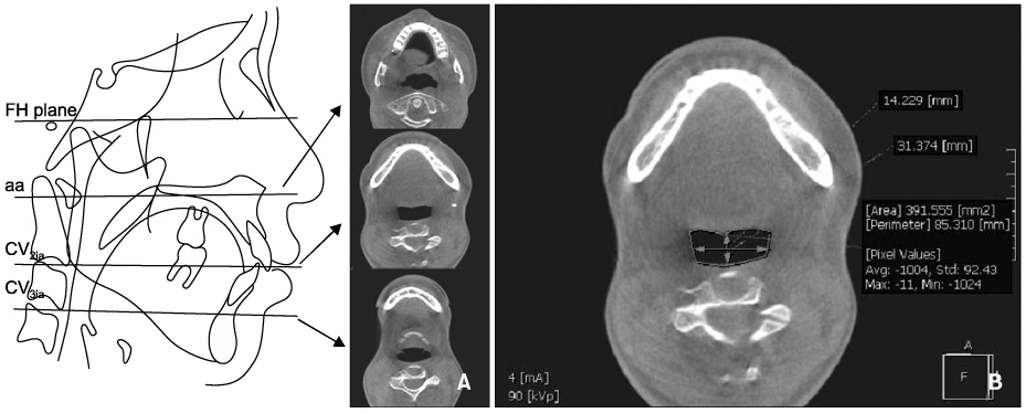
Fig. 1 A, Reference plane (FH plane, aa plane, CV2 plane, CV3 plane); B, measurements of pharyngeal airway width and area on the reference plane.
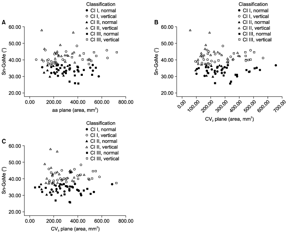
Fig. 2 A, Pharyngeal airway areas of subgroups (vertical and normal pattern) in aa plane; B, pharyngeal airway areas of subgroups (vertical and normal pattern) in CV2 plane; C, pharyngeal airway areas of subgroups (vertical and normal pattern) in CV3 plane.
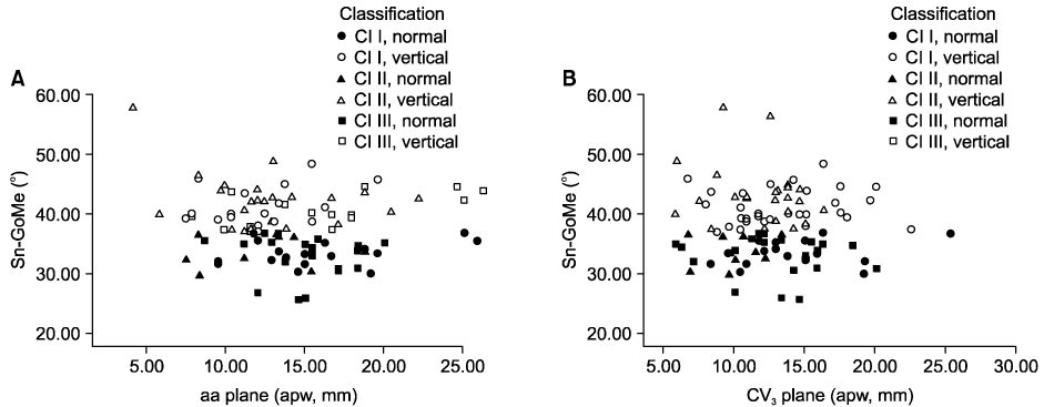
Fig. 3 A, Pharyngeal airway width (apw) of subgroups (vertical and normal pattern) in aa plane; B, pharyngeal airway width (apw) of subgroups (vertical and normal pattern) in CV3 plane.
Cited by 3 articles
-
Three dimensional analysis of the upper airway and facial morphology in children with Class II malocclusion using cone-beam computed tomography
Ji-Suk Hong, Dae-Sung Kim, Kyung-Min Oh, Yoon-Ji Kim, Kyu-Hong Lee, Yang-Ho Park
Korean J Orthod. 2010;40(3):134-144. doi: 10.4041/kjod.2010.40.3.134.Morphological characteristics of the upper airway and pressure drop analysis using 3D CFD in OSA patients
Sung-Seo Mo, Hyung Taek Ahn, Jeong-Seon Lee, Yoo-Sam Chung, Yoon-Shik Moon, Eung-Kwon Pae, Sang-Jin Sung
Korean J Orthod. 2010;40(2):66-76. doi: 10.4041/kjod.2010.40.2.66.Comparison of midsagittal reference plane in PA cephalogram and 3D CT
Jin-Hyoung Cho, Ji-Yeon Moon
Korean J Orthod. 2010;40(1):6-15. doi: 10.4041/kjod.2010.40.1.6.
Reference
-
1. Moss ML, Salentijn L. The primary role of functional matrices in facial growth. Am J Orthod. 1969. 55:566–577.
Article2. Dunn GF, Green LJ, Cunat JJ. Relationship between variation of mandibular morphology and variation of nasopharyngeal airway size in monozygotic twins. Angle Orthod. 1973. 43:129–135.3. Kerr WJ. The nasopharynx, face height, and overbite. Angle Orthod. 1985. 55:31–36.4. McNamara JA Jr. A method of cephalometric evaluation. Am J Orthod. 1984. 86:449–469.
Article5. Hwang YI, Lee KH, Lee KJ, Kim SC, Cho HJ, Cheon SH, et al. Effect of airway and tongue in facial morphology of prepubertal Class I, II children. Korean J Orthod. 2008. 38:74–82.
Article6. Lee YS, Kim JC. A cephalometric study on the airway size according to the types of the malocclusion. Korean J Orthod. 1995. 25:19–20.7. Son WS, Choi YS. Evaluation of hyoid bone position and airway size in Class III malocclusion. Korean J Orthod. 1996. 26:247–254.8. Kwak SY, Kim HY, Jeon YM, Kim JG. Airway size in mlocclusions with hyperdivergent skeletal pattern. Korean J Orthod. 2003. 33:293–305.9. Kyung SH, Park YC, Pae EK. Obstructive sleep apnea patients with the oral appliance experience pharyngeal size and shape changes in three dimensions. Angle Orthod. 2005. 75:15–22.10. Rachmiel A, Aizenbud D, Pillar G, Srouji S, Peled M. Bilateral mandibular distraction for patients with compromised airway analyzed by three-dimensional CT. Int J Oral Maxillofac Surg. 2005. 34:9–18.
Article11. Kim HJ, Park HS, Kwon OW. Evaluation of potency of panoramic radiography for estimating the position of maxillary impacted canines using 3D CT. Korean J Orthod. 2008. 38:265–274.
Article12. Jeon HJ, Park SH, Jung SH, Chun YS. Three dimensional analysis of tooth movement using different sizes of NiTi wire on NiTi scissors-bite corrector. Korean J Orthod. 2009. 39:43–53.
Article13. Hwang SH, Park IS, Nam KY, Kim JB, Cho YW, Suh YS, et al. Cephalometric differences in obstructive sleep apnea between obese and non-obese Korean male patients. Korean J Orthod. 2008. 38:202–213.
Article14. Major MP, Flores-Mir C, Major PW. Assessment of lateral cephalometric diagnosis of adenoid hypertrophy and posterior upper airway obstruction: a systemic review. Am J Orthod Dentofaca Orthop. 2006. 130:700–708.
Article15. Jeans WD, Fernando DC, Maw AR, Leighton BC. A longitudinal study of the growth of the nasopharynx and its contents in normal children. Br J Radiol. 1981. 54:117–121.
Article16. Lagravère MO, Carey J, Toogood RW, Major PW. Three-dimensional accuracy of measurements made with software on cone-beam computed tomography images. Am J Orthod Dentofacial Orthop. 2008. 134:112–116.
Article17. Chang HS, Baik HS. A proposal of landmarks for craniofacial analysis using three-dimensional CT imaging. Korean J Orthod. 2002. 32:313–325.18. Ludlow JB, Davies-Ludlow LE, Brooks SL, Howerton WB. Dosimetry of 3 CBCT devices for oral and maxillofacial radiology: CB Mercurary, NewTom 3G and i-CAT. Dentomaxillofac Radiol. 2006. 35:219–226.
Article19. Danforth RA, Dus I, Mah J. 3-D volume imaging for dentistry: a new dimension. J Calf Dent Assoc. 2003. 31:817–823.20. Anegawa E, Tsuyama H, Kusakawa J. Lateral cephalometric analysis of the pharyngeal airway space affected by head posture. Int J Oral Maxillofac Surg. 2008. 37:805–809.
Article21. de Freitas MR, Alcazar NM, Janson G, de Freitas KM, Henriques JF. Upper and lower pharyngeal airway in subjects with Class I and Class II malocclusions and different growth patterns. Am J Orthod Dentofacial Orthop. 2006. 130:742–745.
Article22. Ceylan I, Okatay H. A study on the pharyngeal size in different skeletal patterns. Am J Orthod Dentofac Orthop. 1995. 108:69–75.
Article23. Chen F, Terada K, Hua Y, Saito I. Effects of bimaxillary surgery and mandibular setback surgery on pharyngeal airway measurements in patients with Class III skeletal deformities. Am J Orthod Dentofacial Orthop. 2007. 131:372–377.
Article
- Full Text Links
-
- Actions
-
Cited
- CITED
-
- Close
- Share
-
- Similar articles
-
- Assessment of pharyngeal airway in Korean adolescents according to skeletal pattern, sex, and cervical vertebral maturation: A cross-sectional CBCT study
- Detection of maxillary second molar with two palatal roots using cone beam computed tomography: a case report
- Can Dental Cone Beam Computed Tomography Assess Bone Mineral Density?
- Three dimensional cone-beam CT study of upper airway change after mandibular setback surgery for skeletal Class III malocclusion patients
- Observation of mandibular second molar roots and root canal morphology using dental cone-beam computed tomography
