Three dimensional cone-beam CT study of upper airway change after mandibular setback surgery for skeletal Class III malocclusion patients
- Affiliations
-
- 1Department of Orthodontics, School of Dentistry, Pusan National University, Korea. sbypark@pusan.ac.kr
- 2Department of Oral & Maxillofacial Surgery, School of Dentistry, Pusan National University, Korea.
- KMID: 1459568
- DOI: http://doi.org/10.4041/kjod.2010.40.3.145
Abstract
OBJECTIVE
Lateral cephalometric radiographs have been the main form of resource for assessing two dimensional anteroposterior airway changes. The purpose of this study was to evaluate the three dimensional volumetric change in the upper airway space in Class III malocclusion patients who underwent mandibular setback surgery.
METHODS
Three dimensional cone-beam computed tomographs (CBCT) and their three dimensional reconstruction images were analyzed. The samples consisted of 20 adult patients (12 males and 8 females) who were diagnosed as skeletal Class III and underwent mandibular setback surgery. CBCTs were taken at 3 stages - Baseline (1.8 weeks before surgery), T1 (2.3 months after surgery), and T2 (1 year after surgery). Pharyngeal airway was separated according to the reference planes and reconstructed into the nasopharynx, the oropharynx and the hypopharynx. Measurements at Baseline, T1, and T2 were compared between groups.
RESULTS
The result showed the volume of the pharyngeal airway decreased significantly 2.3 months after surgery (p < 0.001) and the diminished airway did not recover after 1 year post-surgery. The oropharynx was the most decreased area.
CONCLUSIONS
These findings suggest that mandibular setback surgery causes both short-term and long-term decrease in the upper airway space.
MeSH Terms
Figure
-
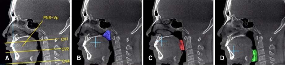
Fig. 1 Three dimensional image and MPR (multiplanar projection reformat) overlay views of airways. A, Reference planes (PNS-Vp plane, CV1 plane, CV2 plane, CV4 plane); B, volumetric image at nasopharynx area; C, volumetric image at oropharynx area; D, volumetric image at hypopharynx area. PNS, Psterior nasal spine; Vp, the most posterior point of the ala of vomer; CV, cervical vertebrae.
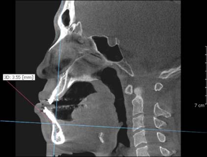
Fig. 2 Measurement of mandibular position. Mandibular position was evaluated using anotomical B point at each time period.
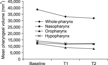
Fig. 3 Mean pharyngeal volume change. Baseline, Before surgery; T1, 2.3 months after surgery; T2, 1 year after surgery.
Cited by 1 articles
-
Three dimensional structural analysis between dental arch and basal bone in normal occlusion
Jee-Tae Kim, Jin-Woo Lee
Korean J Orthod. 2011;41(4):224-236. doi: 10.4041/kjod.2011.41.4.224.
Reference
-
1. Baik HS. Characteristics of craniofacial structures in Class III malocclusion. 1991. Daegu: School of Dentistry, Kyungpook Natioinal University;Master thesis.2. Lee DK, Kim SK. A study on the changes in the upper airway following osteotomy for the mandibular prognathism. J Korean Dent Assoc. 1989. 27:1143–1153.3. King EW. A roentgenographic study of pharyngeal growth. Angle Orthod. 1952. 23–37.4. Brodie AG. Anatomy and physiology of head and neck musculature. Am J Orthod. 1950. 36:831–844.
Article5. Handelman CS, Osborne G. Growth of the nasopharynx and adenoid development from one to eighteeen years. Angle Orthod. 1976. 46:243–259.6. Kollias I, Krogstad O. Adult craniocervical and pharyngeal changes - a longitudinal cephalometric study between 22 and 42 years of age. Part I: Morphological craniocervical and hyoid bone changes. Eur J Orthod. 1999. 21:333–344.
Article7. Johnston CD, Richardson A. Cephalometric changes in adult pharyngeal morphology. Eur J Orthod. 1999. 21:357–362.
Article8. Kim KW, Choung JI, Kim CH. A cephalometric study on changes in hyoid bone, tongue and upper airway space according to skeletal change in persons with mandible prognathism after orthognathic surgery. J Korean Assoc Maxillofac Plast Reconstr Surg. 2004. 26:349–358.9. Chen F, Terada K, Hua Y, Saito I. Effects of bimaxillary surgery and mandibular setback surgery on pharyngeal airway measurements in patients with Class III skeletal deformities. Am J Orthod Dentofacial Orthop. 2007. 131:372–377.
Article10. Athanasiou AE, Toutountzakis N, Mavreas D, Ritzau M, Wenzel A. Alterations of hyoid bone position and pharyngeal depth and their relationship after surgical correction of mandibular prognathism. Am J Orthod Dentofacial Orthop. 1991. 100:259–265.
Article11. Wickwire NA, White RP Jr, Proffit WR. The effect of mandibular osteotomy on tongue position. J Oral Surg. 1972. 30:184–190.12. Tselnik M, Pogrel MA. Assessment of the pharyngeal airway space after mandibular setback surgery. J Oral Maxillofac Surg. 2000. 58:282–285.
Article13. Eggensperger N, Smolka W, Iizuka T. Long-term changes of hyoid bone position and pharyngeal airway size following mandibular setback by sagittal split ramus osteotomy. J Craniomaxillofac Surg. 2005. 33:111–117.
Article14. Hochban W, Schurmann R, Brandenburg U, Conradt R. Mandibular setback for surgical correction of mandibular hyperplasia - does it provoke sleep-related breathing disorders? Int J Oral Maxillofac Surg. 1996. 25:333–338.
Article15. Samman N, Tang SS, Xia J. Cephalometric study of the upper airway in surgically corrected class III skeletal deformity. Int J Adult Orthodon Orthognath Surg. 2002. 17:180–190.16. Enacar A, Aksoy AU, Sencift Y, Haydar B, Aras K. Changes in hypopharyngeal airway space and in tongue and hyoid bone positions following the surgical correction of mandibular prognathism. Int J Adult Orthodon Orthognath Surg. 1994. 9:285–290.17. Kawakami M, Yamamoto K, Fujimoto M, Ohgi K, Inoue M, Kirita T. Changes in tongue and hyoid positions, and posterior airway space following mandibular setback surgery. J Craniomaxillofac Surg. 2005. 33:107–110.
Article18. Lee SH. A study of relapse and position of hyoid bone following orthognathic surgery. J Korean Assoc Maxillofac Plast Reconstr Surg. 1991. 13:476–490.19. Wenzel A, Williams S, Ritzau M. Changes in head posture and nasopharyngeal airway following surgical correction of mandibular prognathism. Eur J Orthod. 1989. 11:37–42.
Article20. Holmberg H, Linder-Anderson S. Cephalometric radiographs as a means of evaluating the capacity of the nasal and nasopharyngeal airway. Am J Orthod. 1979. 76:479–490.
Article21. Chung DH, Lee KS. A study on changes of airway, tongue, and hyoid position following orthognathic surgery. Korean J Orthod. 1998. 28:487–498.22. Baumrind S, Frantz RC. The reliability of head film measurements. 1. Landmark identification. Am J Orthod. 1971. 60:111–127.23. Major MP, Flores-Mir C, Major PW. Assessment of lateral cephalometric diagnosis of adenoid hypertrophy and posterior upper airway obstruction: a systematic review. Am J Orthod Dentofacial Orthop. 2006. 130:700–708.
Article24. Park SH, Yu HS, Kim KD, Lee KJ, Baik HS. A proposal for a new analysis of craniofacial morphology by 3-dimensional computed tomography. Am J Orthod Dentofacial Orthop. 2006. 129:600.e23–600.e34.
Article25. Mah J, Hatcher D. Three-dimensional craniofacial imaging. Am J Orthod Dentofacial Orthop. 2004. 126:308–309.
Article26. Lye KW. Effect of orthognathic surgery on the posterior airway space (PAS). Ann Acad Med Singapore. 2008. 37:677–682.27. Stuck BA, Maurer JT. Airway evaluation in obstructive sleep apnea. Sleep Med Rev. 2008. 12:411–436.
Article28. Kawamata A, Fujishita M, Ariji Y, Ariji E. Three-dimensional computed tomographic evaluation of morphologic airway changes after mandibular setback osteotomy for prognathism. Oral Surg Oral Med Oral Pathol Oral Radiol Endod. 2000. 89:278–287.
Article29. Degerliyurt K, Ueki K, Hashiba Y, Marukawa K, Nakagawa K, Yamamoto E. A comparative CT evaluation of pharyngeal airway changes in class III patients receiving bimaxillary surgery or mandibular setback surgery. Oral Surg Oral Med Oral Pathol Oral Radiol Endod. 2008. 105:495–502.
Article30. Greco JM, Frohberg U, Van Sickels JE. Long-term airway space changes after mandibular setback using bilateral sagittal split osteotomy. Int J Oral Maxillofac Surg. 1990. 19:103–105.
Article31. Lee YS, Baik HS, Lee KJ, Yu HS. The structural change in the hyoid bone and upper airway after orthognathic surgery for skeletal class III anterior open bite patients using 3-dimentional computed tomography. Korean J Orthod. 2009. 39:72–82.
Article32. Joss CU, Vassalli IM. Stability after bilateral sagittal split osteotomy setback surgery with rigid internal fixation: a systematic review. J Oral Maxillofac Surg. 2008. 66:1634–1643.
Article33. Kitagawara K, Kobayashi T, Goto H, Yokobayashi T, Kitamura N, Saito C. Effects of mandibular setback surgery on oropharyngeal airway and arterial oxygen saturation. Int J Oral Maxillofac Surg. 2008. 37:328–333.
Article34. Lowe AA, Fleetham JA, Adachi S, Ryan CF. Cephalometric and computed tomographic predictors of obstructive sleep apnea severity. Am J Orthod Dentofacial Orthop. 1995. 107:589–595.
Article35. Pepin JL, Ferretti G, Veale D, Romand P, Coulomb M, Brambilla C, et al. Somnofluoroscopy, computed tomography, and cephalometry in the assessment of the airway in obstructive sleep apnoea. Thorax. 1992. 47:150–156.
Article36. Riley R, Guilleminault C, Herran J, Powell N. Cephalometric analyses and flow-volume loops in obstructive sleep apnea patients. Sleep. 1983. 6:303–311.
Article
- Full Text Links
-
- Actions
-
Cited
- CITED
-
- Close
- Share
-
- Similar articles
-
- Three-dimensional analysis of pharyngeal airway change of skeletal class III patients in cone beam computed tomography after bimaxillary surgery
- The structural change in the hyoid bone and upper airway after orthognathic surgery for skeletal class III anterior open bite patients using 3-dimensional computed tomography
- Postoperative changes in the pharyngeal airway space through computed tomography evaluation after mandibular setback surgery in skeletal class III patients: 1-year follow-up
- Changes of the hyoid bone position and the upper airway dimension after orthognathic surgery in skeletal class III patients
- Evaluation of hyoid bone position and airway size in Class III malocclusion
