The structural change in the hyoid bone and upper airway after orthognathic surgery for skeletal class III anterior open bite patients using 3-dimensional computed tomography
- Affiliations
-
- 1Department of Orthodontics, College of Dentistry, Yonsei University, Korea.
- 2Department of Orthodontics, College of Dentistry, Oral Science Research Center, The Institute of Cranio-facial Deformity, Yonsei University, Korea. yumichael@yuhs.ac
- KMID: 1975812
- DOI: http://doi.org/10.4041/kjod.2009.39.2.72
Abstract
OBJECTIVE
The purpose of this study was to investigate the structural changes of the hyoid bone and upper airway after orthognathic surgery for skeletal class III anterior open bite patients, and make comparisons with normal occlusion. METHODS: Pre- and post-operative computed tomography (CT) examinations were performed on 12 skeletal class III anterior open bite patients who were treated with mandibular setback osteotomy. Using the V-works 4.0(TM) program, 3-dimensional images of the total skull, mandible, hyoid bone, and upper airway were evaluated. RESULTS: In the Class III openbite group, the hyoid bone were all positioned anteriorly, compared to the Normal group (p < 0.05). The angle between the hyoid plane and mandibular plane in the Class III openbite group before surgery was greater than in the Normal group (p < 0.05), and the difference increased after surgery (p < 0.01). In the Class III openbite group, the volume of the upper airway decreased after surgery (p < 0.001) and the volume of the upper airway was smaller than the Normal group before and after surgery (p < 0.001). CONCLUSIONS: The narrow upper airway space in skeletal Class III openbite patients decreased after mandibular setback osteotomy. This may affect the post-surgical stability.
Keyword
Figure
-

Fig 1 Procedure of 3D image reconstruction using V-works™ 4.0 (Cybermed, Seoul, Korea).
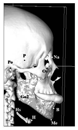
Fig 2 Nine landmarks used in this study. Na (Nasion), Most posterior point on the curvature between frontal bone and nasal bone in the midsagittal plane; P (prechiasmatic groove), vertical and transverse midpoint of prechiasmatic groove; Or (orbitale), lowest point on the infraorbital margin of each orbit; Po (porion, anatomical), highest midpoint on the roof of the external auditory meatus; B (B point), greatest concavity point on the anterior border of the symphysis; Me (menton), most inferior point on the symphysis of the mandible; Go2 (gonion2), midpoint of the posterior border of mandibular angle; H (hyoidale), most upper and superior point at the middle of the hyoid bone; Hs (hyoidale superioris), most upper and posterior point at the greater horn of hyoid bone.
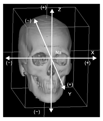
Fig 3 Coordinate axis used in this study. A positive coordinate value indicates the front, superior, and left side of the patient, and a negative value indicates the opposite. Na (Nasion), Most posterior point on the curvature between frontal bone and nasal bone in the midsagittal plane.

Fig 4 Measurements of Hyoid bone. The 3-dimensional position of hyoid bone was obtained by measuring the distances between H point and Coronal, Midsagittal, and Horizontal planes. The angle between hyoid plane and horizontal, mandibular planes was obtained for the angular measurement. If the angle between hyoid plane and mandibular plane was more than 180°, a negative value was used and defined as reverse inclination. Five reference planes were used in this study. Horizontal plane, parallel to the FH plane, which was constructed on both sides of Po and left of Or, passing through Na; Midsagittal plane, perpendicular to the horizontal plane passing through Na and P; Coronal plane, at right angles to the horizontal and midsagittal plane passing through Na; Mandibular plane, constructed by Me and both sides of Go2; Hyoid plane, constructed by H and both sides of Hs.
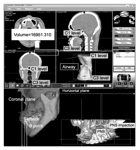
Fig 5 Measurements of Airway space, Mandibular setback, PNS impaction. 3-dimensional reconstruction of upper airway was limited between C1 and C3. Mandibular setback was obtained by measuring the difference in the distance between Coronal plane and B point at pre-, post-surgery. PNS impaction was obtained by measuring the difference in the distance between Horizontal plane and PNS point at pre-, post-surgery. C1, lowest midpoint on the 1st cervical vertebrae; C3, lowest midpoint on the 3rd cervical vertebrae; B (B point), greatest concavity point on the anterior border of the symphysis; PNS (posterior nasal spine), the process formed by uniting the projecting ends of the posterior borders of the palatal process of the palatal bone; sBack, difference of mandibular set back at T1 and T2; pIMPACTION, difference of PNS.
Cited by 2 articles
-
Three dimensional analysis of the upper airway and facial morphology in children with Class II malocclusion using cone-beam computed tomography
Ji-Suk Hong, Dae-Sung Kim, Kyung-Min Oh, Yoon-Ji Kim, Kyu-Hong Lee, Yang-Ho Park
Korean J Orthod. 2010;40(3):134-144. doi: 10.4041/kjod.2010.40.3.134.Three dimensional cone-beam CT study of upper airway change after mandibular setback surgery for skeletal Class III malocclusion patients
Na-Ri Kim, Yong-Il Kim, Soo-Byung Park, Dae-Seok Hwang
Korean J Orthod. 2010;40(3):145-155. doi: 10.4041/kjod.2010.40.3.145.
Reference
-
1. Cho IJ, Rhee BT. A cephalometric study on the position of the hyoid bone in cleft lip and palate individuals. Korean J Orthod. 1990. 20:197–207.2. Seo BM, Min BI. Skeletal relapse after sagittal split osteotomies for correction of mandibular prognathism. J Korean Oral Maxillofac Surg. 1991. 17:32–39.3. Katakura N, Umino M, Kubota Y. Morphologic airway changes after mandibular setback osteotomy for prognathism with and without cleft palate. Anesth Pain Control Dent. 1993. 2:22–26.4. Greco JM, Frohberg U, Van Sickels JE. Long-term airway space changes after mandibular setback using bilateral sagittal split osteotomy. Int J Oral Maxillofac Surg. 1990. 19:103–105.
Article5. Paik UB, Lee HC. A study of change of tongue position, hyoid bone position, and upper airway following sagittal split ramus osteotomy in mandible prognathism patients. Inje Med J. 1999. 20:457–466.6. Moss ML, Salentijn L. The primary role of functional matrices in facial growth. Am J Orthod. 1969. 55:566–577.
Article7. Chung HS, Lee KS. The effect of functional pressures of the tongue and lips on the incisor relationship. Korean J Orthod. 1983. 13:15–30.8. Cuozzo GS, Bowman DC. Hyoid positioning during deglutition following forced positioning of the tongue. Am J Orthod. 1975. 68:564–570.
Article9. Tallgren A, Solow B. Hyoid bone position, facial morphology and head posture in adults. Eur J Orthod. 1987. 9:1–8.
Article10. Park SH, Yu HS, Kim KD, Lee KJ, Baik HS. A proposal for a new analysis of craniofacial morphology by 3-dimensional computed tomography. Am J Orthod Dentofacial Orthop. 2006. 129:600.e23–600.e34.
Article11. Cavalcanti MG, Vannier MW. Quantitative analysis of spiral computed tomography for craniofacial clinical applications. Dentomaxillofac Radiol. 1998. 27:344–350.
Article12. Kim HJ, Park HS, Kwon OW. Evaluation of potency of panoramic radiography for estimating the position of maxillary impacted canines using 3D CT. Korean J Orthod. 2008. 38:265–274.
Article13. Jeon YN, Lee KH, Hwang HS. Validity of midsagittal reference planes constructed in 3D CT images. Korean J Orthod. 2007. 37:182–191.14. Kawamata A, Fujishita M, Ariji Y, Ariji E. Three-dimensional computed tomographic evaluation of morphologic airway changes after mandibular setback osteotomy for prognathism. Oral Surg Oral Med Oral Pathol Oral Radiol Endod. 2000. 89:278–287.
Article15. Wickwire NA, White RP Jr, Proffit WR. The effect of mandibular osteotomy on tongue position. J Oral Surg. 1972. 30:184–190.16. Swanson LT, Murray JE. Partial glossectomy to stabilize occlusion following surgical correction of prognathism. Report of a case. Oral Surg Oral Med Oral Pathol. 1969. 27:707–715.
Article17. Enacar A, Aksoy AU, Sençift Y, Haydar B, Aras K. Changes in hypopharyngeal airway space and in tongue and hyoid bone positions following the surgical correction of mandibular prognathism. Int J Adult Orthodon Orthognath Surg. 1994. 9:285–290.18. King EW. A roentgenographic study of pharyngeal growth. Angle Orthod. 1952. 22:23–37.19. Adamidis IP, Spyropoulos MN. Hyoid bone position and orientation in Class I and Class III malocclusions. Am J Orthod Dentofacial Orthop. 1992. 101:308–312.
Article20. Song YJ, Kim HJ, Nam SH, Kim YJ. Hyoid bone position in Class I, II and III malocclusions. J Korean Acad Pediatr Dent. 1999. 26:564–571.21. Chang YI. A radiographic study of the hyoid bone position in malocclusion. Korean J Orthod. 1987. 17:7–14.22. Chin KS, Shon WS. The relationships between the postoperative stability and the changes in the tongue position, thehyoid bone position and the upper airway size after orthognathic surgery in patients with mandibular prognathism. Korean J Orthod. 1993. 23:693–706.23. Athanasiou AE, Toutountzakis N, Mavreas D, Ritzau M, Wenzel A. Alterations of hyoid bone position and pharyngeal depth and their relationship after surgical correction of mandibular prognathism. Am J Orthod Dentofacial Orthop. 1991. 100:259–265.
Article24. Gu G, Gu G, Nagata J, Suto M, Anraku Y, Nakamura K, et al. Hyoid position, pharyngeal airway and head posture in relation to relapse after the mandibular setback in skeletal Class III. Clin Orthod Res. 2000. 3:67–77.
Article25. Tallgren A, Solow B. Long-term changes in hyoid bone position and craniocervical posture in complete denture wearers. Acta Odontol Scand. 1984. 42:257–267.
Article26. Ferrario VF, Sforza C, Germanò D, Dalloca LL, Miani A Jr. Head posture and cephalometric analyses: an integrated photographic/ radiographic technique. Am J Orthod Dentofacial Orthop. 1994. 106:257–264.
Article27. Stepovich ML. A cephalometric positional study of the hyoid bone. Am J Orthod. 1965. 51:882–900.
Article28. Wenzel A, Williams S, Ritzau M. Changes in head posture and nasopharyngeal airway following surgical correction of mandibular prognathism. Eur J Orthod. 1989. 11:37–42.
Article29. Santos Junior JF, Abrahão M, Gregório LC, Zonato AI, Gumieiro EH. Genioplasty for genioglossus muscle advancement in patients with obstructive sleep apnea-hypopnea syndrome and mandibular retrognathia. Braz J Otorhinolaryngol. 2007. 73:480–486.
Article30. Heller JB, Gabbay JS, Kwan D, O'Hara CM, Garri JI, Urrego A, et al. Genioplasty distraction osteogenesis and hyoid advancement for correction of upper airway obstruction in patients with Treacher Collins and Nager syndromes. Plast Reconstr Surg. 2006. 117:2389–2398.
Article31. Kerr WJ. The nasopharynx, face height, and overbite. Angle Orthod. 1985. 55:31–36.32. Dunn GF, Green LJ, Cunat JJ. Relationships between variation of mandibular morphology and variation of nasopharyngeal airway size in monozygotic twins. Angle Orthod. 1973. 43:129–135.33. O'Ryan FS, Gallagher DM, LaBanc JP, Epker BN. The relation between nasorespiratory function and dentofacial morphology: a review. Am J Orthod. 1982. 82:403–410.34. Takagi Y, Gamble JW, Proffit WR, Christiansen RL. Postural change of the hyoid bone following osteotomy of the mandible. Oral Surg Oral Med Oral Pathol. 1967. 23:688–692.
Article35. Eggensperger N, Smolka W, Iizuka T. Long-term changes of hyoid bone position and pharyngeal airway size following mandibular setback by sagittal split ramus osteotomy. J Craniomaxillofac Surg. 2005. 33:111–117.
Article36. Kawakami M, Yamamoto K, Fujimoto M, Ohgi K, Inoue M, Kirita T. Changes in tongue and hyoid positions, and posterior airway space following mandibular setback surgery. J Craniomaxillofac Surg. 2005. 33:107–110.
Article
- Full Text Links
-
- Actions
-
Cited
- CITED
-
- Close
- Share
-
- Similar articles
-
- A study on relation of position of hyoidbone and upper airway dimensional change according to chin movement in persons with skeletal class III facial pattern after orthognathic surgery
- Changes of the hyoid bone position and the upper airway dimension after orthognathic surgery in skeletal class III patients
- Evaluation of hyoid bone position and airway size in Class III malocclusion
- The three dimensional analysis of volumetric airway change in orthognathic surgery of mandibular prognathism
- Change of the upper airway after mandibular setback surgery in patients with mandibular prognathism and anterior open bite
