Korean J Leg Med.
2012 May;36(1):85-96. 10.7580/KoreanJLegMed.2012.36.1.85.
Application of 3D Surface Scanners in Forensic Science and Medicine ( I ): Digital Storage of Human Skeletons and Development of Appraisal Methods for Incident Scenes
- Affiliations
-
- 1Korea Disaster Victim Identification, National Forensic Service, Seoul, Korea. nechung@korea.kr
- 2Division of Forensic Medicine, National Forensic Service, Seoul, Korea.
- 3Division of Questioned Document, National Forensic Service, Seoul, Korea.
- 4Division of Traffic Engineering, National Forensic Service, Seoul, Korea.
- 5Division of Physical Analysis, National Forensic Service, Seoul, Korea.
- 6Division of Forensic Medicine, the Central District Office of National Forensic Service, Daejon, Korea.
- 7Department of Anatomy, College of Medicine, Soonchunhyang University, Cheonan, Korea.
- 8Department of Anatomy, Ewha Womans University School of Medicine, Seoul, Korea.
- 9Department of Anatomy/Catholic Institute for Applied Anatomy, College of Medicine, Catholic University, Seoul, Korea.
- 10Aviation and Railway Accident Investigation Board, Seoul, Korea.
- 11Cadastral Research Institute of Korea Cadastral Survey Corporation, Seoul, Korea.
- 12Opto Mechanical Associate Co., Daejon, Korea.
- 13Dimension & Solution Co., Daejon, Korea.
- 14Dream Technology & Solution Co., Seongnam, Korea.
- 15Wipco Co., Seongnam, Korea.
- 16Multimedia Business & System Provider Co. Seoul, Korea.
- KMID: 2250323
- DOI: http://doi.org/10.7580/KoreanJLegMed.2012.36.1.85
Abstract
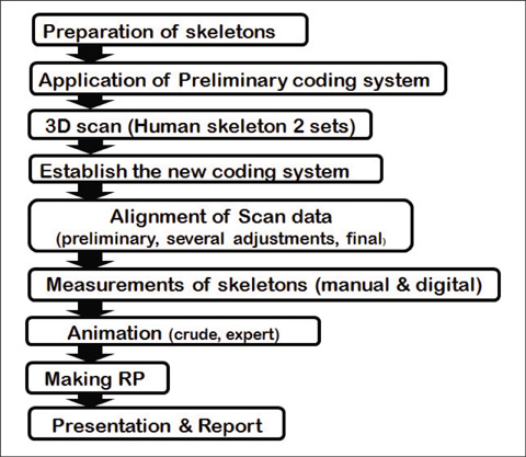
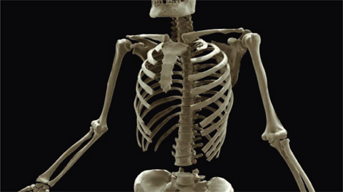
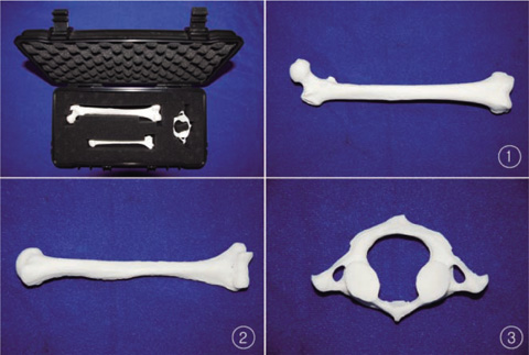
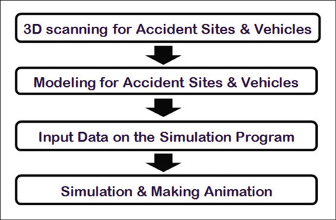
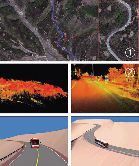
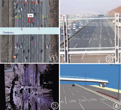
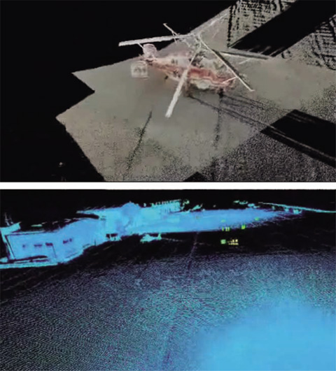
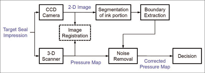
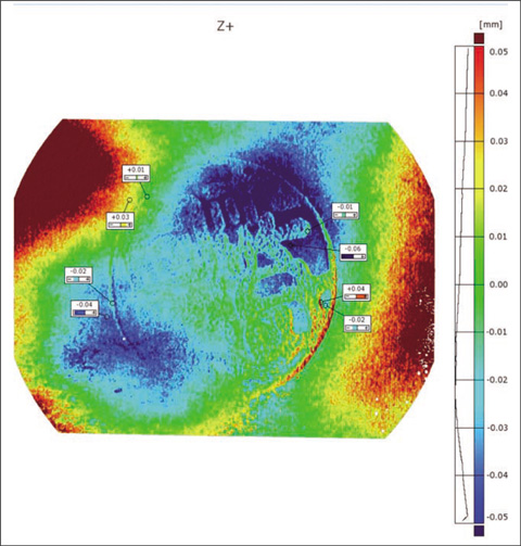
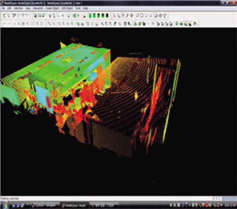
- The aim of this project was to use 3D scanning data collected at incident scenes and various evidence to 1) develop surveying methods based on 3D data consisting of overall and detailed scene evidence, captured by long-range and micros-canner, which can be shared by personnel working in different fields such as forensic medicine, video analysis, physical analysis, traffic engineering, and fire investigation; 2) create digital storage for human skeletons and set the foundation for virtual anthropology; and 3) improve the credibility of 3D evidence by virtual remodeling and simulation of incident scenes and evidence to provide a basis for advanced and high-tech scientific investigation. Two complete skeletons of male and female were scanned using 3D micro-scanner. Each bone was successfully reproduced and assembled in virtual space. In addition, recreating evidence scheduled for invasive examination by creating RP (rapid prototype) was possible. These outcomes could play an important role in setting up the new field of virtual anthropology. Case-specific surveying methods were developed through analysis of 3D scanning data collected by long-range surface scanners at the scenes of vehicular accidents, falls, shootings, and violent crimes. A technique and recording method was also developed for detecting forged seals by micro-scanning the pressure exerted on the seal. Appraisal methods developed in this project could be utilized to secure 3D data of human skeletal remains and incident scenes, create a standard for application, and increase objectivity, reproducibility, and accuracy of scanning methods. We plan to develop case-specific 3D data analysis techniques to improve the credibility of analysis at the NFS and to establish a 3D data collection and analysis team.
Keyword
MeSH Terms
Figure
Reference
-
1. Chen F, Brown GM, Song M. Overview of three-dimensional shape measurement using optical method. Opt Eng. 2000. 39:10–22.2. Wang YF, Cheng DI. Three-dimensional shape construction and recognition by fusing intensity and structured lighting. J Pattern Rec Soc. 1992. 25:1411–1425.3. O'Neill M, Denos M. Automated system for coarse-to-fine pyramidal area correlation stereo matching. Image and Vision Comput. 1996. 14:220–236.4. Buck U, Naether S, Braun M, et al. Application of 3D documentation and geometric reconstruction methods in traffic accident analysis: With high resolution surface scanning, radiological MSCT/MRI scanning and real data based animation. Forensic Sci Int. 2007. 170:20–28.5. Brüschweiler W, Brauna M, Dirnhoferb R, Thali MJ. Analysis of patterned injuries and injury-causing instruments with forensic 3D/CAD supported photogrammetry (FPHG): an instruction manual for the documentation process. Forensic Sci Int. 2003. 132:130–138.6. Thali MJ, Braunb M, Markwaldera H, et al. Bite mark documentation and analysis: the forensic 3D/CAD supported photogrammetry approach. Forensic Sci Int. 2003. 135:115–121.7. Lee J, Lee ED, Tark HO, et al. Study of photogrammetric comparison method of patterned injuries using 3D CAD program. Korean J Leg Med. 2004. 28:32–37.8. Yang KM, Chung NE, Hong SW, et al. Preliminary research for applying appraisal techniques to evaluate the cause of skin injury. Korean J Leg Med. 2008. 32:105–110.9. Lee J, Lee ED, Tark HO, Hwang JW, Yoon DY. Efficient height measurement method of surveillance camera image. Forensic Sci Int. 2008. 177:17–23.10. Yoshino M, Taniguchia M, Imaizumia K, et al. A new retrieval system for a database of 3D facial images. Forensic Sci Int. 2005. 148:113–120.11. Ross AH, Williams S. Testing repeatability and error of coordinate landmark data acquired from crania. J Forensic Sci. 2008. 53:782–785.12. Choi JY, Choi JH, Kim NK, et al. Analysis of errors in medical rapid prototyping models. Int J Oral Maxillofac Surg. 2002. 31:23–32.13. Buikstra JE, Ublelaker DH. Standards for data collection from human skeletal remains. 1997. 3rd ed. Akansas: Archeological Survey;67–84.14. Bookstein FL. Morphometric tools for landmark data. 1991. Cambridge: Cambridge University Press;63–66.15. Slice DE. Modern morphometrics in physical anthropology. 2005. New York: Kluwer Academic / Plenum Publishers;8–10.16. Rohr K. Fundamental limits in 3D landmark localization. Inf Process Med Imaging. 2005. 19:286–298.17. Forsberg A, Kullberg J, Agartz I, Ahlstrom H, Johansson L, Henriksson KM. Landmark-based software for anatomical measurements: a precision study. Clin Anat. 2009. 22:456–462.18. Subburaj K, Ravi B, Agarwal M. Automated identification of anatomical landmarks on 3D bone models reconstructed from CT scan images. Comput Med Imaging Graph. 2009. 33:359–368.
- Full Text Links
- Actions
-
Cited
- CITED
-
- Close
- Share
- Similar articles
-
- A Surface Treatment Technique for Interim Crown Fabricated by Three-Dimensional Printing with Digital Light-Processing Technology
- Bioarchaeological Studies on Ancient Human Skeletons in Korea
- 3D Facial Scanners: How to Make the Right Choice for Orthodontists
- Principles and Fallacies in Medical Appraisal: Focusing on Gastroenterology Cases
- Surface Functionalization of Three-Dimensional Printed Scaffold for Biomedical Application

