J Gynecol Oncol.
2012 Jul;23(3):153-158. 10.3802/jgo.2012.23.3.153.
Tumor volume and lymphovascular space invasion as a prognostic factor in early invasive adenocarcinoma of the cervix
- Affiliations
-
- 1Department of Obstetrics and Gynecology, Keio University School of Medicine, Tokyo, Japan. fujiit@a5.keio.jp
- 2Department of Pathology, Keio University School of Medicine, Tokyo, Japan.
- KMID: 2245173
- DOI: http://doi.org/10.3802/jgo.2012.23.3.153
Abstract
OBJECTIVE
The aim of this study was to investigate the risk and recurrence of early invasive adenocarcinoma of the cervix, and to determine whether non-radical methods of management could be performed.
METHODS
The medical and histopathological records of 50 patients with early invasive adenocarcinoma of the cervix treated at Keio University Hospital between 1993 and 2005 were reviewed, and compared with the literature.
RESULTS
The median follow-up period was 64.3 months. The depth of stromal invasion was < or =3 mm in 33 cases and >3 mm, but < or =5 mm in 17 cases. The horizontal spread was < or =7 mm in 25 cases and >7 mm in 25 cases. One of the 33 cases that had tumor volumes of < or =500 mm3, and three of the 17 cases with tumor volumes of >500 mm3 were positive for lymph node metastasis. When our data were combined with previously reported results, statistically significant differences were observed between the tumor volume and the frequency of pelvic lymph node metastasis/the rate of recurrence (p<0.0001). The frequency of pelvic lymph node metastases was significantly higher in the lymphovascular space invasion (LVSI)-positive group than in the LVSI-negative group (p=0.02). No adnexal metastasis or parametrial involvement was noted.
CONCLUSION
Assessment of the depth of stromal invasion, tumor volume, and LVSI is critical for selecting an appropriate therapeutic modality. Non-radical methods of management are considered suitable for patients with LVSI-negative adenocarcinoma of the cervix exhibiting a stromal invasion depth of < or =5 mm and a tumor volume of < or =500 mm3.
Keyword
MeSH Terms
Figure
-
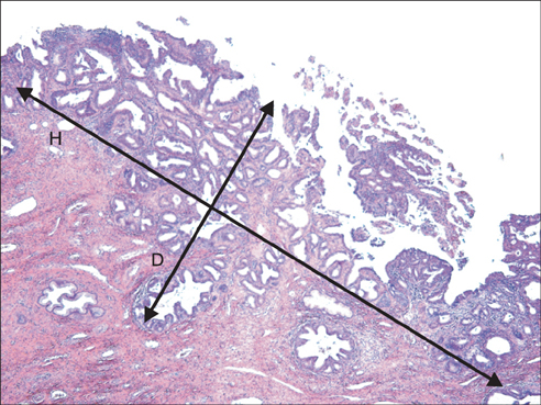
Fig. 1 Diagram of tumor volume measurement calculated using the Burghardt method. Tumor volume was determined using the following equation: depth (D) of stromal invasion×horizontal (H) spread×1.5 greater value of depth or horizontal spread.
Reference
-
1. DiSaia PJ, Creasman WT. Clinical gynecologic oncology. 1997. 5th ed. St. Louis: Mosby.2. Jones WB, Mercer GO, Lewis JL Jr, Rubin SC, Hoskins WJ. Early invasive carcinoma of the cervix. Gynecol Oncol. 1993. 51:26–32.3. McGonigle KF, Berek JS. Early-stage squamous cell and adenocarcinoma of the cervix. Curr Opin Obstet Gynecol. 1992. 4:109–119.4. Matsukuma K, Tsukamoto N, Kaku T, Matsumura M, Toki N, Toh N, et al. Early adenocarcinoma of the uterine cervix - its histologic and immunohistologic study. Gynecol Oncol. 1989. 35:38–43.5. Rollason TP, Cullimore J, Bradgate MG. A suggested columnar cell morphological equivalent of squamous carcinoma in situ with early stromal invasion. Int J Gynecol Pathol. 1989. 8:230–236.6. Kaspar HG, Dinh TV, Doherty MG, Hannigan EV, Kumar D. Clinical implications of tumor volume measurement in stage I adenocarcinoma of the cervix. Obstet Gynecol. 1993. 81:296–300.7. Kaku T, Kamura T, Sakai K, Amada S, Kobayashi H, Shigematsu T, et al. Early adenocarcinoma of the uterine cervix. Gynecol Oncol. 1997. 65:281–285.8. Ostor AG. Early invasive adenocarcinoma of the uterine cervix. Int J Gynecol Pathol. 2000. 19:29–38.9. Kurian K, al-Nafussi A. Relation of cervical glandular intraepithelial neoplasia to microinvasive and invasive adenocarcinoma of the uterine cervix: a study of 121 cases. J Clin Pathol. 1999. 52:112–117.10. Nicklin JL, Perrin LC, Crandon AJ, Ward BG. Microinvasive adenocarcinoma of the cervix. Aust N Z J Obstet Gynaecol. 1999. 39:411–413.11. Elliott P, Coppleson M, Russell P, Liouros P, Carter J, MacLeod C, et al. Early invasive (FIGO stage IA) carcinoma of the cervix: a clinico-pathologic study of 476 cases. Int J Gynecol Cancer. 2000. 10:42–52.12. Covens A, Kirby J, Shaw P, Chapman W, Franseen E. Prognostic factors for relapse and pelvic lymph node metastases in early stage I adenocarcinoma of the cervix. Gynecol Oncol. 1999. 74:423–427.13. Webb JC, Key CR, Qualls CR, Smith HO. Population-based study of microinvasive adenocarcinoma of the uterine cervix. Obstet Gynecol. 2001. 97(5 Pt 1):701–706.14. Schorge JO, Lee KR, Flynn CE, Goodman A, Sheets EE. Stage IA1 cervical adenocarcinoma: definition and treatment. Obstet Gynecol. 1999. 93:219–222.15. Schorge JO, Lee KR, Sheets EE. Prospective management of stage IA(1) cervical adenocarcinoma by conization alone to preserve fertility: a preliminary report. Gynecol Oncol. 2000. 78:217–220.16. Burghardt E. Microinvasive carcinoma in gynaecological pathology. Clin Obstet Gynaecol. 1984. 11:239–257.17. Kasamatsu T, Okada S, Tsuda H, Shiromizu K, Yamada T, Tsunematsu R, et al. Early invasive adenocarcinoma of the uterine cervix: criteria for nonradical surgical treatment. Gynecol Oncol. 2002. 85:327–332.18. Bisseling KC, Bekkers RL, Rome RM, Quinn MA. Treatment of microinvasive adenocarcinoma of the uterine cervix: a retrospective study and review of the literature. Gynecol Oncol. 2007. 107:424–430.19. Nagarsheth NP, Maxwell GL, Bentley RC, Rodriguez G. Bilateral pelvic lymph node metastases in a case of FIGO stage IA(1) adenocarcinoma of the cervix. Gynecol Oncol. 2000. 77:467–470.20. Utsugi K, Shimizu Y, Akiyama F, Hasumi K. Is the invasion depth in millimeters valid to determine the prognosis of early invasive cervical adenocarcinoma? A case of recurrent FIGO stage IA1 cervical adenocarcinoma. Gynecol Oncol. 2001. 82:205–207.21. Smith HO, Qualls CR, Romero AA, Webb JC, Dorin MH, Padilla LA, et al. Is there a difference in survival for IA1 and IA2 adenocarcinoma of the uterine cervix? Gynecol Oncol. 2002. 85:229–241.22. Hirai Y, Takeshima N, Tate S, Akiyama F, Furuta R, Hasumi K. Early invasive cervical adenocarcinoma: its potential for nodal metastasis or recurrence. BJOG. 2003. 110:241–246.23. Balega J, Michael H, Hurteau J, Moore DH, Santiesteban J, Sutton GP, et al. The risk of nodal metastasis in early adenocarcinoma of the uterine cervix. Int J Gynecol Cancer. 2004. 14:104–109.24. Ceballos KM, Shaw D, Daya D. Microinvasive cervical adenocarcinoma (FIGO stage 1A tumors): results of surgical staging and outcome analysis. Am J Surg Pathol. 2006. 30:370–374.25. Chung CK, Nahhas WA, Stryker JA, Curry SL, Abt AB, Mortel R. Analysis of factors contributing to treatment failures in stages IB and IIA carcinoma of the cervix. Am J Obstet Gynecol. 1980. 138:550–556.26. Boyce J, Fruchter RG, Nicastri AD, Ambiavagar PC, Reinis MS, Nelson JH Jr. Prognostic factors in stage I Carcinoma of the cervix. Gynecol Oncol. 1981. 12(2 Pt 1):154–165.27. van Nagell JR Jr, Donaldson ES, Wood EG, Parker JC Jr. The significance of vascular invasion and lymphocytic infiltration in invasive cervical cancer. Cancer. 1978. 41:228–234.28. Abdulhayoglu G, Rich WM, Reynolds J, DiSaia PJ. Selective radiation therapy in stage IB uterine cervical carcinoma following radical pelvic surgery. Gynecol Oncol. 1980. 10:84–92.29. Boyce JG, Fruchter RG, Nicastri AD, DeRegt RH, Ambiavagar PC, Reinis M, et al. Vascular invasion in stage I carcinoma of the cervix. Cancer. 1984. 53:1175–1180.30. Fujii T, Nakamura M, Kameyama K, Saito M, Nishio H, Ohno A, et al. Digital colposcopy for the diagnosis of cervical adenocarcinoma using a narrow band imaging system. Int J Gynecol Cancer. 2010. 20:605–610.
- Full Text Links
-
- Actions
-
Cited
- CITED
-
- Close
- Share
-
- Similar articles
-
- Multivariate prognostic analysis of adenocarcinoma of the uterine cervix treated with radical hysterectomy and systematic lymphadenectomy
- Clinical Significance of Tumor Angiogenesis in Squamous Cell Carcinoma of the Uterine Cervix
- Clinicopathologic characteristics and prognostic factors in adenocarcinoma of the uterine cervix
- E - cadherin Expression in Carcinoma of The Uterine Cervix
- A Histopathological Analysis of 69 Cases of Adenocarcinoma of the Uterine Cervix
