The Effect of Radiographic Contrast Media on Reperfusion Injury in the Isolated Rat Heart
- Affiliations
-
- 1Department of Internal Medicine, Pusan National University Yangsan Hospital, Yangsan, Korea.
- 2Institute of Cancer Rehabilitation and Convalescence, Yoonsung Hospital, Cheongdo, Korea.
- 3Department of Preventive Medicine, Keimyung University School of Medicine, Daegu, Korea.
- 4Division of Cardiology, Department of Internal Medicine and Research Institute for Convergence of Biomedical Science and Technology, Pusan National University Yangsan Hospital, Pusan National University School of Medicine, Yangsan, Korea. junehongk@gmail.
- 5Cardiovascular Research Laboratory, Pusan National University Yangsan Hospital, Yangsan, Korea.
- KMID: 2223859
- DOI: http://doi.org/10.4070/kcj.2014.44.6.423
Abstract
- BACKGROUND AND OBJECTIVES
We investigated the effects of commonly used contrast media (CM) on myocardial ischemia-reperfusion injury in isolated rat hearts.
SUBJECTS AND METHODS
Isolated rat hearts were subjected to 30 minutes of regional ischemia and 2 hours of reperfusion. The following CM (1 mL/1 L Krebs-Henseleit buffer) were randomly perfused for 15 minutes beginning 5 minutes before reperfusion and ending 10 minutes after reperfusion: iohexol (n=8), iopromide (n=8), ioversol (n=8), iomeprol (n=8), iopamidol (n=7), ioxaglate (n=8), and iodixanol (n=7). The effects of a direct bolus injection of undiluted iohexol, iopromide, or ioxaglate (each n=6) via the aortic root immediately prior to reperfusion were also evaluated. The area of necrosis, expressed as the percentage of the area at risk (AN/AR), and cardiodynamic variables were measured.
RESULTS
The AN/AR of the control and experimental groups in the order described in methods was 33.7+/-6.4%, 30.3+/-7.4%, 34.7+/-12.6%, 29.2+/-10.2%, 20.9+/-7.6%, 22.6+/-8.7%, 18.8+/-7.9%, and 19.9+/-11.4%, respectively. Groups that received iomeprol and ioxaglate exhibited significantly decreased AN/AR values compared to those of control hearts (p=0.042 and p=0.013). No significant differences in the AN/AR were observed between control hearts and the groups injected with a single bolus of CM. No significant hemodynamic changes were noted after reperfusion among the groups.
CONCLUSION
The overall effects of the CM on coronary reperfusion were not deleterious, and better effects were noted in two CM groups. However, it is unclear whether this result was attributed to a specific physiochemical property of the CM.
MeSH Terms
Figure
-
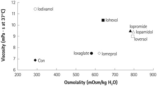
Fig. 1 Physiochemical properties of the contrast media (CM), particularly focusing on osmolality and viscosity. Note that ioxaglate 320 and iodixanol 320 have similar viscosity and osmolality.
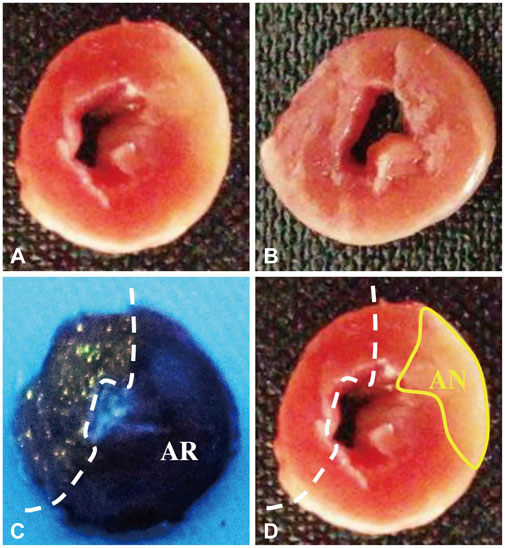
Fig. 2 Representative images obtained during measurements. The midportion of the left ventricle of a control heart (A) and a diluted ioxaglateperfused heart (B) after triphenyltetrazolium chloride (TTC) staining are shown. Intact myocardium can be discriminated by the fluorescent area under UV light. The other side of the dotted line is the area at risk (AR) (C). The area of necrosis (AN) is identified as the region not stained by TTC under natural light (D). UV: ultraviolet.
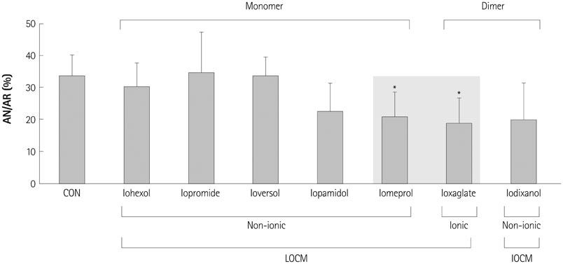
Fig. 3 Area of necrosis (AN) as a percentage of the area at risk (AR) in protocol 1. Contrast media (CM) was perfused continuously from 5 minutes before reperfusion to 10 minutes after reperfusion. Shaded box indicates the lower viscosity group. Values are means±standard deviations *p<0.05 vs. CON. CON: untreated control hearts, LOCM: low-osmolality CM, IOCM: iso-osmolality CM.
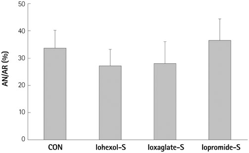
Fig. 4 Area of necrosis (AN) as a percentage of the area at risk (AR) in protocol 2. Iohexol-S, iopromide-S, and ioxaglate-S indicate single bolus injections of iohexol, iopromide, and ioxaglate, respectively. Values are means± standard deviations. No significant differences in AN/AR were observed between the CON and experimental groups. CON: untreated control hearts.
Reference
-
1. Heinrich MC, Kuhlmann MK, Grgic A, Heckmann M, Kramann B, Uder M. Cytotoxic effects of ionic high-osmolar, nonionic monomeric, and nonionic iso-osmolar dimeric iodinated contrast media on renal tubular cells in vitro. Radiology. 2005; 235:843–849.2. Aspelin P, Aubry P, Fransson SG, et al. Nephrotoxic effects in high-risk patients undergoing angiography. N Engl J Med. 2003; 348:491–499.3. Romano G, Briguori C, Quintavalle C, et al. Contrast agents and renal cell apoptosis. Eur Heart J. 2008; 29:2569–2576.4. Wessely R, Koppara T, Bradaric C, et al. Choice of contrast medium in patients with impaired renal function undergoing percutaneous coronary intervention. Circ Cardiovasc Interv. 2009; 2:430–437.5. Oriyanhan W, Yamazaki K, Miwa S, Takaba K, Ikeda T, Komeda M. Taurine prevents myocardial ischemia/reperfusion-induced oxidative stress and apoptosis in prolonged hypothermic rat heart preservation. Heart Vessels. 2005; 20:278–285.6. Gonzalez-Castillo C, Rubio R, Zenteno-Savin T. Coronary flow-induced inotropism is modulated by binding of dextrans to the endothelial luminal surface. Am J Physiol Heart Circ Physiol. 2003; 284:H1348–H1357.7. Seeliger E, Sendeski M, Rihal CS, Persson PB. Contrast-induced kidney injury: mechanisms, risk factors, and prevention. Eur Heart J. 2012; 33:2007–2015.8. Sendeski MM. Pathophysiology of renal tissue damage by iodinated contrast media. Clin Exp Pharmacol Physiol. 2011; 38:292–299.9. Seeliger E, Becker K, Ladwig M, Wronski T, Persson PB, Flemming B. Up to 50-fold increase in urine viscosity with iso-osmolar contrast media in the rat. Radiology. 2010; 256:406–414.10. Falck G, Bruvold M, Schjøtt J, Jynge P. Protective effects of repetitive injections of radiographic contrast media on the subsequent tolerance to ischemia in the isolated rat heart. Cardiovasc Intervent Radiol. 2000; 23:466–471.11. Piper HM, García-Dorado D. Prime causes of rapid cardiomyocyte death during reperfusion. Ann Thorac Surg. 1999; 68:1913–1919.12. Falck G, Schjott J, Jynge P. Hyperosmotic pretreatment reduces infarct size in the rat heart. Physiol Res. 1999; 48:331–340.13. Pastukh V, Ricci C, Solodushko V, Mozaffari M, Schaffer SW. Contribution of the PI 3-kinase/Akt survival pathway toward osmotic preconditioning. Mol Cell Biochem. 2005; 269:59–67.14. Cecchi E, Liotta AA, Gori AM, et al. Relationship between blood viscosity and infarct size in patients with ST-segment elevation myocardial infarction undergoing primary percutaneous coronary intervention. Int J Cardiol. 2009; 134:189–194.15. Wasilewski J, Turczyński B, Słowińska L, Kowalik V, Osadnik T, Poloński L. Haemorheological factors and myocardial reperfusion in patients with ST-elevation myocardial infarction undergoing primary coronary intervention. Kardiol Pol. 2007; 65:778–785. discussion 786-7.16. Yalcin O, Ulker P, Yavuzer U, Meiselman HJ, Baskurt OK. Nitric oxide generation by endothelial cells exposed to shear stress in glass tubes perfused with red blood cell suspensions: role of aggregation. Am J Physiol Heart Circ Physiol. 2008; 294:H2098–H2105.17. Schmid I, Didier D, Pfammatter T, et al. Effects of non-ionic iodinated contrast media on patient heart rate and pressures during intra-cardiac or intra-arterial injection. Int J Cardiol. 2007; 118:389–396.18. Schmiedel E. Evaluation of the adverse effects of iomeprol. Eur J Radiol. 1994; 18:Suppl 1. S104–S108.19. Reed M, Meier P, Tamhane UU, Welch KB, Moscucci M, Gurm HS. The relative renal safety of iodixanol compared with low-osmolar contrast media: a meta-analysis of randomized controlled trials. JACC Cardiovasc Interv. 2009; 2:645–654.20. Heinrich MC, Häberle L, Müller V, Bautz W, Uder M. Nephrotoxicity of iso-osmolar iodixanol compared with nonionic low-osmolar contrast media: meta-analysis of randomized controlled trials. Radiology. 2009; 250:68–86.21. From AM, Al Badarin FJ, McDonald FS, Bartholmai BJ, Cha SS, Rihal CS. Iodixanol versus low-osmolar contrast media for prevention of contrast induced nephropathy: meta-analysis of randomized, controlled trials. Circ Cardiovasc Interv. 2010; 3:351–358.
- Full Text Links
-
- Actions
-
Cited
- CITED
-
- Close
- Share
-
- Similar articles
-
- Effect of Ischemic Preconditioning on Catecholamine Release from the Isolated, Ischemic Reperfused Hearts of Rats
- Experimental Studies on the Effect of Ginsenoside Rg1 Mixtures in an Isolated Rat Heart after Ischemic Arrest and Reperfusion
- The effects of hydrogen sulfide under sevoflurane administration against ischemia and reperfusion injury in isolated rat heart
- Effects of postconditioning with N,N,N'N'-tetrakis-[2-pyridylmethyl]-ethylenediamine in isolated rat hearts
- Effect of C1 esterase inhibitor on the cardiac dysfunction following ischemia and reperfusion in the isolated perfused rat heart
