Korean Circ J.
2014 Nov;44(6):406-414. 10.4070/kcj.2014.44.6.406.
Usefulness of the Doppler Flow of the Ophthalmic Artery in the Evaluation of Carotid and Coronary Atherosclerosis
- Affiliations
-
- 1Department of Cardiology, Daegu Catholic University Medical Center, Daegu, Korea. kks7379@cu.ac.kr
- KMID: 2223857
- DOI: http://doi.org/10.4070/kcj.2014.44.6.406
Abstract
- BACKGROUND AND OBJECTIVES
There is little information about the relationship between the Doppler flow of the ophthalmic artery (OA) and carotid and coronary atherosclerosis. The aim of the investigation was to assess the clinical usefulness of the Doppler flow of the OA to estimate the severity of carotid and coronary atherosclerosis.
SUBJECTS AND METHODS
The study was a retrospective analysis of the findings in 140 patients (mean age: 60 years, male: 64%) who underwent coronary angiography (CA) for the evaluation of typical angina between July 2010 and October 2011 in our single center. The severity of coronary artery stenosis was based on the Gensini score (GS). Significant coronary artery disease (CAD) was defined as the obstruction of over 75% of the major coronary arteries confirmed with CA. The pulsed Doppler flow of the OA and carotid ultrasound were performed before CA.
RESULTS
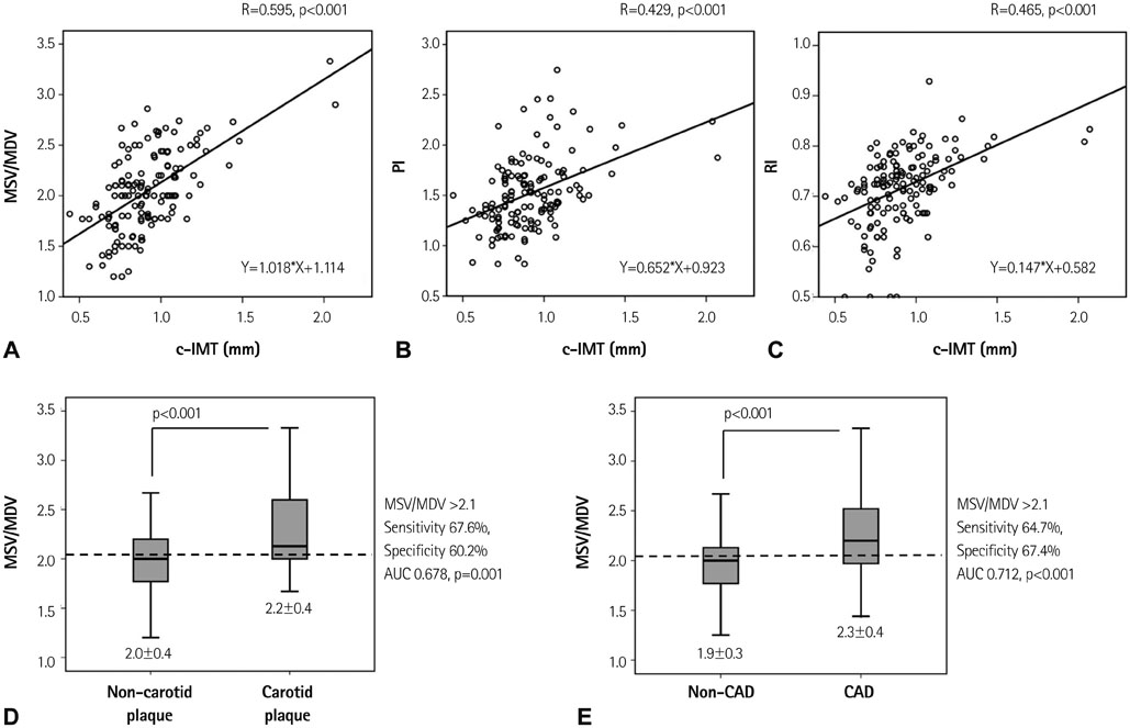
The mean systolic velocity/mean diastolic velocity (MSV/MDV), pulsatile index and resistance index in the Doppler flow of the OA were identified as significant and independent correlations with carotid intima-media thickness, and MSV/MDV was identified to have a significant and independent correlation with the GS. MSV/MDV >2.1 was the independent predictor for significant CAD {odds ratio (OR) 3.8, 95% confidence interval (CI) 1.5-9.7, p=0.005} and carotid plaque (OR 2.8, 95% CI 1.1-7.0, p=0.028), after adjustment for CAD-associated factors.
CONCLUSION
The Doppler flow of the OA might be a useful predictor of the severity of carotid and coronary atherosclerosis.
MeSH Terms
Figure
Reference
-
1. Burger-Kentischer A, Göbel H, Kleemann R, et al. Reduction of the aortic inflammatory response in spontaneous atherosclerosis by blockade of macrophage migration inhibitory factor (MIF). Atherosclerosis. 2006; 184:28–38.2. Stein JH, Korcarz CE, Hurst RT, et al. Use of carotid ultrasound to identify subclinical vascular disease and evaluate cardiovascular disease risk: a consensus statement from the American Society of Echocardiography Carotid Intima-Media Thickness Task Force. Endorsed by the Society for Vascular Medicine. J Am Soc Echocardiogr. 2008; 21:93–111. quiz 189-90.3. Lorenz MW, Markus HS, Bots ML, Rosvall M, Sitzer M. Prediction of clinical cardiovascular events with carotid intima-media thickness: a systematic review and meta-analysis. Circulation. 2007; 115:459–467.4. Zielinski T, Dzielinska Z, Januszewicz A, et al. Carotid intima-media thickness as a marker of cardiovascular risk in hypertensive patients with coronary artery disease. Am J Hypertens. 2007; 20:1058–1064.5. Ralph RA. Prediction of cardiovascular status from arteriovenous crossing phenomena. Ann Ophthalmol. 1974; 6:323–326.6. Michelson EL, Morganroth J, Nichols CW, MacVaugh H 3rd. Retinal arteriolar changes as an indicator of coronary artery disease. Arch Intern Med. 1979; 139:1139–1141.7. Gillum RF. Retinal arteriolar findings and coronary heart disease. Am Heart J. 1991; 122(1 Pt 1):262–263.8. Wong TY, Klein R, Klein BE, Tielsch JM, Hubbard L, Nieto FJ. Retinal microvascular abnormalities and their relationship with hypertension, cardiovascular disease, and mortality. Surv Ophthalmol. 2001; 46:59–80.9. Tedeschi-Reiner E, Strozzi M, Skoric B, Reiner Z. Relation of atherosclerotic changes in retinal arteries to the extent of coronary artery disease. Am J Cardiol. 2005; 96:1107–1109.10. Maruyoshi H, Kojima S, Kojima S, et al. Waveform of ophthalmic artery Doppler flow predicts the severity of systemic atherosclerosis. Circ J. 2010; 74:1251–1256.11. Hu HH, Sheng WY, Yen MY, Lai ST, Teng MM. Color Doppler imaging of orbital arteries for detection of carotid occlusive disease. Stroke. 1993; 24:1196–1203.12. European Society of Hypertension-European Society of Cardiology Guidelines Committee. 2003 European Society of Hypertension-European Society of Cardiology guidelines for the management of arterial hypertension. J Hypertens. 2003; 21:1011–1053.13. ADA Statement. Diagnosis and classification of diabetes mellitus. Diabetes Care. 2013; 36:67–74.14. Expert Panel on Detection, Evaluation, and Treatment of High Blood Cholesterol in Adults. Executive Summary of The Third Report of The National Cholesterol Education Program (NCEP) Expert Panel on Detection, Evaluation, And Treatment of High Blood Cholesterol In Adults (Adult Treatment Panel III). JAMA. 2001; 285:2486–2497.15. Gensini GG. A more meaningful scoring system for determining the severity of coronary heart disease. Am J Cardiol. 1983; 51:606.16. Lieb WE, Cohen SM, Merton DA, Shields JA, Mitchell DG, Goldberg BB. Color Doppler imaging of the eye and orbit. Technique and normal vascular anatomy. Arch Ophthalmol. 1991; 109:527–531.17. Yatsuka Y, Matsukubo S, Tsutsumi K, Kotegawa T, Nakamura K, Nakano S. Ophthalmic artery flow velocity after physical exercise in healthy men. Jpn J Ophthalmol. 1996; 40:103–110.18. Metz CE, Herman BA, Shen JH. Maximum likelihood estimation of receiver operating characteristic (ROC) curves from continuously-distributed data. Stat Med. 1998; 17:1033–1053.19. Tranquart F, Bergès O, Koskas P, et al. Color Doppler imaging of orbital vessels: personal experience and literature review. J Clin Ultrasound. 2003; 31:258–273.20. National Cholesterol Education Program (NCEP) Expert Panel on Detection, Evaluation, and Treatment of High Blood Cholesterol in Adults (Adult Treatment Panel III). Third Report of the National Cholesterol Education Program (NCEP) Expert Panel on Detection, Evaluation, and Treatment of High Blood Cholesterol in Adults (Adult Treatment Panel III) final report. Circulation. 2002; 106:3143–3421.21. Pearson TA, Blair SN, Daniels SR, et al. AHA Guidelines for Primary Prevention of Cardiovascular Disease and Stroke: 2002 Update: Consensus Panel Guide to Comprehensive Risk Reduction for Adult Patients Without Coronary or Other Atherosclerotic Vascular Diseases. American Heart Association Science Advisory and Coordinating Committee. Circulation. 2002; 106:388–391.22. Chobanian AV, Bakris GL, Black HR, et al. Seventh report of the Joint National Committee on Prevention, Detection, Evaluation, and Treatment of High Blood Pressure. Hypertension. 2003; 42:1206–1252.23. Sarnak MJ, Levey AS, Schoolwerth AC, et al. Kidney disease as a risk factor for development of cardiovascular disease: a statement from the American Heart Association Councils on Kidney in Cardiovascular Disease, High Blood Pressure Research, Clinical Cardiology, and Epidemiology and Prevention. Circulation. 2003; 108:2154–2169.24. Grundy SM, Cleeman JI, Merz CN, et al. Implications of recent clinical trials for the National Cholesterol Education Program Adult Treatment Panel III guidelines. Circulation. 2004; 110:227–239.25. Dzau VJ, Antman EM, Black HR, et al. The cardiovascular disease continuum validated: clinical evidence of improved patient outcomes: part I: Pathophysiology and clinical trial evidence (risk factors through stable coronary artery disease). Circulation. 2006; 114:2850–2870.26. Matthiessen ET, Zeitz O, Richard G, Klemm M. Reproducibility of blood flow velocity measurements using colour decoded Doppler imaging. Eye (Lond). 2004; 18:400–405.27. Németh J, Kovács R, Harkányi Z, Knézy K, Sényi K, Marsovszky I. Observer experience improves reproducibility of color Doppler sonography of orbital blood vessels. J Clin Ultrasound. 2002; 30:332–335.28. Päivänsalo M, Pelkonen O, Rajala U, Keinanen-Kiukaanniemi S, Suramo I. Diabetic retinopathy: sonographically measured hemodynamic alterations in ocular, carotid, and vertebral arteries. Acta Radiol. 2004; 45:404–410.29. Fukuda M, Suzuki A, Okamoto N, Tsubakimori S. Investigation of Doppler ultrasound waveform indices in the ophthalmic artery: comparison between results in normal and diabetic eyes. Folia Ophthalmol Jpn. 1999; 50:218–223.30. Päivänsalo M, Riiheläinen K, Rissanen T, Suramo I, Laatikainen L. Effect of an internal carotid stenosis on orbital blood velocity. Acta Radiol. 1999; 40:270–275.
- Full Text Links
- Actions
-
Cited
- CITED
-
- Close
- Share
- Similar articles
-
- General principles of carotid Doppler ultrasonography
- Relationship Between Carotid and Coronary Atherosclerosis in the Elderly
- Carotid ultrasound in patients with coronary artery disease
- Neutrophil-to-Lymphocyte Ratio for Risk Assessment in Coronary Artery Disease and Carotid Artery Atherosclerosis
- Usefulness of External Carotid Artery Angiogram with Manual Carotid Compression in Ophthalmic Artery Aneurysm


