J Cerebrovasc Endovasc Neurosurg.
2019 Jun;21(2):94-100. 10.7461/jcen.2019.21.2.94.
Usefulness of External Carotid Artery Angiogram with Manual Carotid Compression in Ophthalmic Artery Aneurysm
- Affiliations
-
- 1Department of Neurosurgery, Daegu Catholic University Hospital, Catholic University College of Medicine, Daegu, Korea. fhjhcho@cu.ac.kr
- KMID: 2465202
- DOI: http://doi.org/10.7461/jcen.2019.21.2.94
Abstract
OBJECTIVE
Identifying collaterals from external carotid artery (ECA) is necessary before treatment of ophthalmic artery (OphA) aneurysm. We present a manual carotid compression test to verify collaterals in ophthalmic artery aneurysms, and evaluate its usefulness.
MATERIALS AND METHODS
From March 2013 to December 2017, endovascular coiling was performed 19 consecutive patients with 20 OphA aneurysms. We performed manual carotid compression test for patients who had aneurysms incorporating entry of OphA. Clinical and angiographic outcomes were investigated.
RESULTS
Of 13 cases underwent manual carotid compression test, 12 cases were confirmed collateral flow from ECA to OphA. During the coil embolization, we tried to maintain the original OphA flow even if it has a collateral anastomosis. Among them, OphA occlusion occurred in one patient during coiling. Recurrence of aneurysm was occurred in a ruptured case and additional embolization was required.
CONCLUSIONS
The manual carotid compression test is useful method to identify the collaterals from ECA in patients with OphA aneurysm. This test can be used as a screening test for confirming collateral flow in OphA aneurysms or as an alternative for patients who are difficult to perform BTO.
MeSH Terms
Figure
-
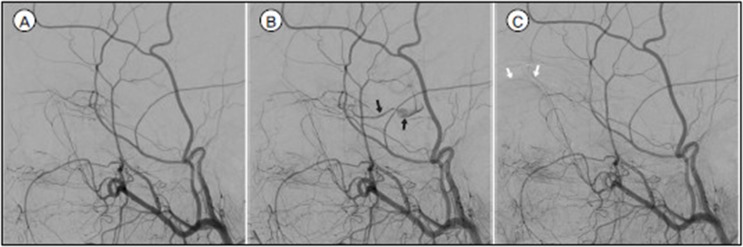
Fig. 1 A. In routine left ECA angiography, there was no flow to OphA. B. and C. During a manual carotid compression test, retrograde blood flow from ECA to OphA (black arrows) and choroidal blush (white arrows) appeared.
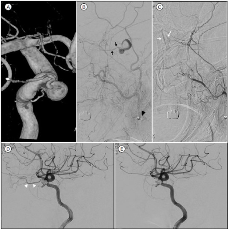
Fig. 2 A. 3-dimensional imaging of DSA of a 65-year-old woman (Case 7) revealed an OphA aneurysm with incorporating the origin of OphA. B. Black arrowhead shows the distal tip of angiographic catheter was located as far away as possible beyond the facial artery. B. and C. Manual carotid compression test confirmed retrograde blood flow from ECA (black arrows) and choroidal blush (white arrows). D. and E. The OphA (white arrowheads) was occluded after coil embolization.
Reference
-
1. Ahn JH, Cho YD, Kang HS, Kim JE, Cho WS, Jung SC, et al. Endovascular treatment of ophthalmic artery aneurysms: assessing balloon test occlusion and preservation of vision in coil embolization. AJNR Am J Neuroradiol. 2014; Nov-Dec. 35(11):2146–2152. PMID: 24970549.
Article2. Andaluz N, Tomsick TA, Keller JT, Zuccarello M. Subdural hemorrhage in the posterior fossa caused by a ruptured cavernous carotid artery aneurysm after a balloon occlusion test. Case report. J Neurosurg. 2006; 8. 105(2):315–319.3. Geibprasert S, Pongpech S, Armstrong D, Krings T. Dangerous extracranial-intracranial anastomoses and supply to the cranial nerves: vessels the neurointerventionalist needs to know. AJNR Am J Neuroradiol. 2009; 9. 30(8):1459–1468. PMID: 19279274.
Article4. Hayreh SS. Orbital vascular anatomy. Eye (Lond). 2006; 10. 20(10):1130–1144. PMID: 17019411.
Article5. Hongo H, Inoue T, Tamura A, Saito I. Surgical strategy to minimize ischemia during trapping/resection of giant extracranial carotid artery aneurysm stratified by collateral evaluation. Surg Neurol Int. 2017; 8:28. PMID: 28303208.
Article6. Kai Y, Morioka M, Yano S, Nakamura H, Makino K, Takeshima H, et al. External Manual Carotid Compression is Effective in Patients with Cavernous Sinus Dural Arteriovenous Fistulae. Interventional Neuroradiology. 2007; 3. 13 Suppl 1:115–122. PMID: 20566088.
Article7. Kim B, Jeon P, Kim K, Yang N, Kim S, Kim H, et al. Endovascular treatment of unruptured ophthalmic artery aneurysms: clinical usefulness of the balloon occlusion test in predicting vision outcomes after coil embolization. J Neurointerv Surg. 2016; 7. 8(7):696–701. PMID: 26113563.
Article8. Lesley WS, Bieneman BK, Dalsania HJ. Selective Use of the Paraophthalmic Balloon Test Occlusion (BTO) to Identify a False-Negative Subset of the Cervical Carotid BTO. Minim Invasive Neurosurg. 2006; 49(1):34–36. PMID: 16547880.
Article9. Linskey ME, Jungreis CA, Yonas H, Hirsch WL Jr, Sekhar LN, Horton JA, et al. Stroke risk after abrupt internal carotid artery sacrifice: accuracy of preoperative assessment with balloon test occlusion and stable xenon-enhanced CT. AJNR Am J Neuroradiol. 1994; 5. 15(5):829–843. PMID: 8059649.10. Lo A, Oehley M, Bartlett A, Adams D, Blyth P, Al-Ali S. Anatomical variations of the common carotid artery bifurcation. ANZ J Surg. 2006; 11. 76(11):970–972. PMID: 17054544.
Article11. Louw L. Different ophthalmic artery origins: Embryology and clinical significance. Clin Anat. 2015; 7. 28(5):576–583. PMID: 25255996.
Article12. Lucev N, Bobinac D, Maric I, Drescik I. Variations of the great arteries in the carotid triangle. Otolaryngol Head Neck Surg. 2000; 4. 122(4):590–591. PMID: 10740186.
Article13. Mathis JM, Barr JD, Jungreis CA, Yonas H, Sekhar LN, Vincent D, et al. Temporary balloon test occlusion of the internal carotid artery: experience in 500 cases. AJNR Am J Neuroradiol. 1995; 4. 16(4):749–754. PMID: 7611033.14. Powers WJ. Cerebral hemodynamics in ischemic cerebrovascular disease. Ann Neurol. 1991; 3. 29(3):231–240. PMID: 2042939.
Article15. Roy D, Milot G, Raymond J. Endovascular treatment of unruptured aneurysms. Stroke. 2001; 9. 32(9):1998–2004. PMID: 11546888.
Article16. van Everdingen KJ, Visser GH, Klijn CJ, Kappelle LJ, van der Grond J. Role of collateral flow on cerebral hemodynamics in patients with unilateral internal carotid artery occlusion. Ann Neurol. 1998; 8. 44(2):167–176. PMID: 9708538.
- Full Text Links
-
- Actions
-
Cited
- CITED
-
- Close
- Share
-
- Similar articles
-
- Aneurysm and Infundibular Dilatation at an Unusual Origin of the Ophthalmic Artery
- Ruptured Aneurysm of the Ophthalmic Artery
- Strategy & Pitfalls of Internal Carotid Artery Aneurysm Surgery
- Extradural-Intradural Approach to Carotid-Ophthalmic Artery Aneurysm
- Intracranial Aneurysm Associated with Aplasia of the Internal Cartoid Artery
