An Assay of Measuring Platelet Reactivity Using Monoclonal Antibody against Activated Platelet Glycoprotein IIb/IIIa in Patients Taking Clopidogrel
- Affiliations
-
- 1Department of Cardiology, Jeju National University Hospital, Jeju, Korea. sejjoo@jejunu.ac.kr
- 2Department of Laboratory Medicine, Jeju National University Hospital, Jeju, Korea.
- KMID: 2223801
- DOI: http://doi.org/10.4070/kcj.2015.45.5.378
Abstract
- BACKGROUND AND OBJECTIVES
Residual platelet reactivity in patients who are taking clopidogrel is commonly measured with VerifyNow assay, which is based on the principle of light transmission aggregometry. However, to evaluate the residual platelet reactivity, it would be more accurate if the reactivity of platelet glycoprotein (GP) IIb/IIIa is directly monitored. In this study, PAC1, a monoclonal antibody against activated platelet GP IIb/IIIa, was used to measure the residual platelet reactivity.
SUBJECTS AND METHODS
Twenty seven patients with coronary artery disease taking clopidogrel were enrolled. Platelets in whole blood were stained with fluorescein isothiocyanate (FITC)-conjugated PAC1. Mean fluorescence intensity (MFI) and % positive platelets (PP) were measured with flow cytometry, and the binding index (BI; MFI x %PP/100) was calculated. P2Y12 reaction unit (PRU) and % inhibition of VerifyNow assay were also measured in the usual manner.
RESULTS
PRU of VerifyNow assay correlated significantly with MFI, %PP, and BI at 10 microM (r=0.59, 0.73, and 0.60, respectively, all p<0.005) and 20 microM of adenosine diphosphate (ADP; r=0.61, 0.75, and 0.63, respectively, all p<0.005). The % inhibition also correlated significantly with MFI, %PP, and BI at 10 microM (r=-0.60, -0.69, and -0.59, respectively, all p<0.005) and 20 microM of ADP (r=-0.63, -0.71, and -0.62, respectively, all p<0.005).
CONCLUSION
Direct measurements of the reactivity of platelet GP IIb/IIIa were feasible using PAC1 and flow cytometry in patients taking clopidogrel. Further clinical studies are required to determine the cut-off values which would define high residual platelet reactivity in patients on this treatment protocol.
MeSH Terms
Figure
-
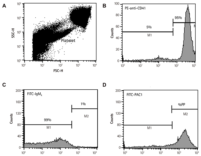
Fig. 1 Flow cytometric analysis of whole blood. (A) Platelets were separated from erythrocytes and white blood cells, on the basis of their characteristic forward- and side- scatter profiles, and a narrow gate was placed around the platelets. (B) Staining of resting platelets with PE-conjugated anti-CD41, a monoclonal antibody against glycoprotein IIb. (C) Staining of platelets with control FITC-conjugated mouse IgMκ. (D) Staining of platelets with FITC-conjugated PAC1. Antibody-positive platelets were defined as those with a fluorescence intensity >99.0% to 99.5%, of non-stimulated platelets stained with FITC-conjugated mouse IgM κ. SSC: side-scatter, FSC: forward-scatter, FITC: fluorescein isothiocyanate, PP: positive platelet.
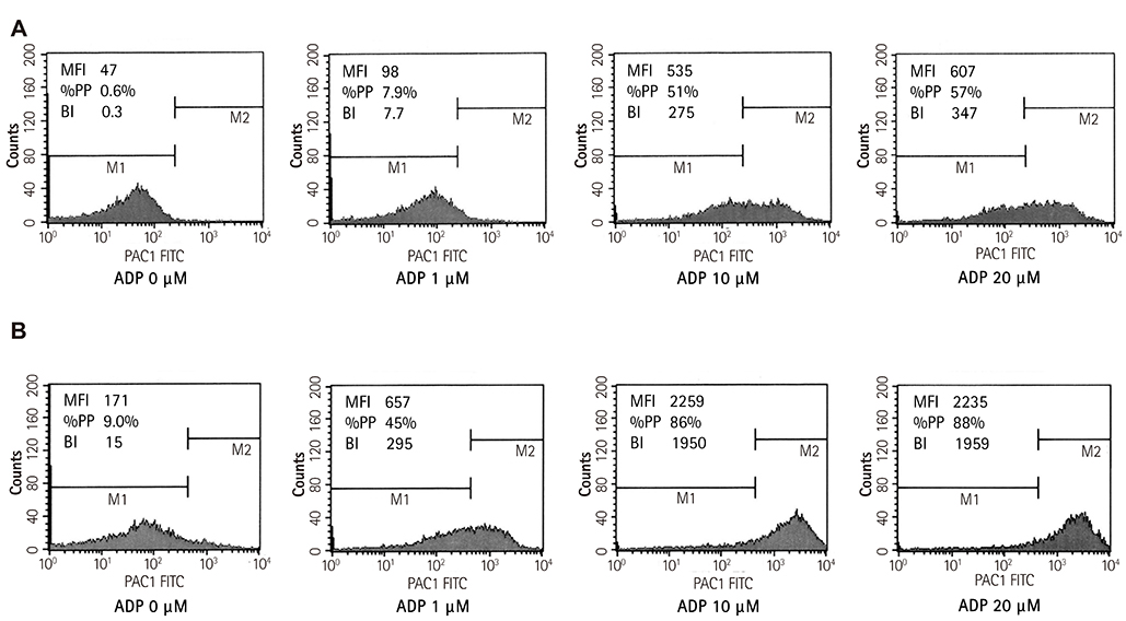
Fig. 2 Examples of flow cytometric data and analysis in a patient with (A) low residual platelet reactivity (B) high residual platelet reactivity. A patient with high residual platelet reactivity showed much greater mean fluorescence intensity (MFI), % positive platelets (%PP), and binding index (BI) at 10 µM or 20 µM of ADP. ADP: adenosine diphosphate, FITC: fluorescein isothiocyanate.
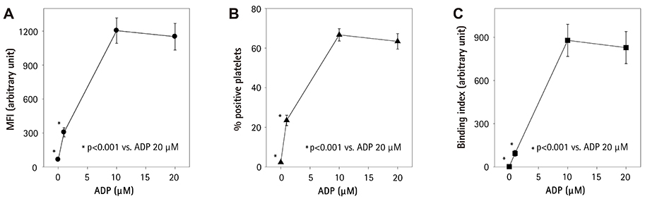
Fig. 3 Changes of mean fluorescence intensity (MFI), % positive platelets, and binding index after ADP stimulation. Values are mean±standard error of mean. They reached maximal values at 10 µM concentration. ADP: adenosine diphosphate.
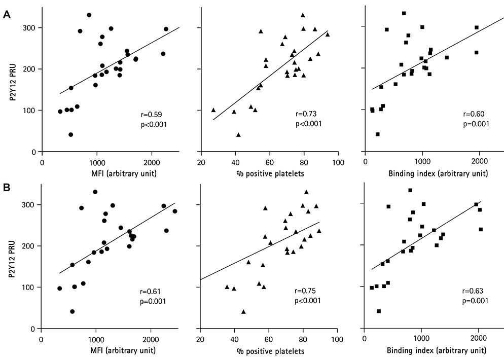
Fig. 4 Correlation between mean fluorescence intensity (MFI), % positive platelets, and binding index with VerifyNow PRU at (A) 10 µM or (B) 20 µM of ADP. PRU: P2Y12 reaction unit, ADP: adenosine diphosphate.
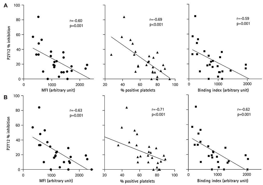
Fig. 5 Correlation between mean fluorescence intensity (MFI), % positive platelets, and binding index with VerifyNow P2Y12 % inhibition at (A) 10 µM or (B) 20 µM of ADP. ADP: adenosine diphosphate.
Reference
-
1. Amsterdam EA, Wenger NK, Brindis RG, et al. 2014 AHA/ACC guideline for the management of patients with non-ST-elevation acute coronary syndromes: executive summary: a report of the American College of Cardiology/American Heart Association task force on practice guidelines. Circulation. 2014; 130:2354–2394.2. O'Gara PT, Kushner FG, Ascheim DD, et al. 2013 ACCF/AHA guideline for the management of ST-elevation myocardial infarction: executive summary: a report of the American College of Cardiology Foundation/American Heart Association task force on practice guidelines. Circulation. 2013; 127:529–555.3. Levine GN, Bates ER, Blankenship JC, et al. 2011 ACCF/AHA/SCAI guideline for percutaneous coronary intervention: executive summary: a report of the American College of Cardiology Foundation/American Heart Association task force on practice guidelines and the Society for Cardiovascular Angiography and Interventions. Circulation. 2011; 124:2574–2609.4. Gurbel PA, Becker RC, Mann KG, Steinhubl SR, Michelson AD. Platelet function monitoring in patients with coronary artery disease. J Am Coll Cardiol. 2007; 50:1822–1834.5. Kuliczkowski W, Witkowski A, Polonski L, et al. Interindividual variability in the response to oral antiplatelet drugs: a position paper of the Working Group on antiplatelet drugs resistance appointed by the Section of Cardiovascular Interventions of the Polish Cardiac Society, endorsed by the Working Group on Thrombosis of the European Society of Cardiology. Eur Heart J. 2009; 30:426–435.6. Brar SS, ten Berg J, Marcucci R, et al. Impact of platelet reactivity on clinical outcomes after percutaneous coronary intervention. A collaborative meta-analysis of individual participant data. J Am Coll Cardiol. 2011; 58:1945–1954.7. Michelson AD. Platelet function testing in cardiovascular diseases. Circulation. 2004; 110:e489–e493.8. Geiger J, Teichmann L, Grossmann R, et al. Monitoring of clopidogrel action: comparison of methods. Clin Chem. 2005; 51:957–965.9. Price MJ. Bedside evaluation of thienopyridine antiplatelet therapy. Circulation. 2009; 119:2625–2632.10. Breet NJ, van Werkum JW, Bouman HJ, et al. Comparison of platelet function tests in predicting clinical outcome in patients undergoing coronary stent implantation. JAMA. 2010; 303:754–762.11. Quinn MJ, Byzova TV, Qin J, Topol EJ, Plow EF. Integrin alphaIIbbeta3 and its antagonism. Arterioscler Thromb Vasc Biol. 2003; 23:945–952.12. Joo SJ. Mechanisms of platelet activation and integrin αIIβ3. Korean Circ J. 2012; 42:295–301.13. Shattil SJ, Hoxie JA, Cunningham M, Brass LF. Changes in the platelet membrane glycoprotein IIb. IIIa complex during platelet activation. J Biol Chem. 1985; 260:11107–11114.14. Shattil SJ, Cunningham M, Hoxie JA. Detection of activated platelets in whole blood using activation-dependent monoclonal antibodies and flow cytometry. Blood. 1987; 70:307–315.15. Abrams CS, Ellison N, Budzynski AZ, Shattil SJ. Direct detection of activated platelets and platelet-derived microparticles in humans. Blood. 1990; 75:128–138.16. Harder S, Klinkhardt U, Graff J, et al. In vitro dose response to different GPIIb/IIIa-antagonists: inter-laboratory comparison of various platelet function tests. Thromb Res. 2001; 102:39–48.17. Furman MI, Kereiakes DJ, Krueger LA, et al. Quantification of abciximab-induced platelet inhibition is assay dependent: a comparative study in patients undergoing percutaneous coronary intervention. Am Heart J. 2003; 145:e6.18. Furman MI, Krueger LA, Linden MD, Barnard MR, Frelinger AL 3rd, Michelson AD. Release of soluble CD40L from platelets is regulated by glycoprotein IIb/IIIa and actin polymerization. J Am Coll Cardiol. 2004; 43:2319–2325.19. Sibbing D, Byrne RA, Bernlochner I, Kastrati A. High platelet reactivity and clinical outcome - fact and fiction. Thromb Haemost. 2011; 106:191–202.20. Price MJ, Berger PB, Teirstein PS, et al. Standard- vs high-dose clopidogrel based on platelet function testing after percutaneous coronary intervention: the GRAVITAS randomized trial. JAMA. 2011; 305:1097–1105.21. Xiao Z, Théroux P. Clopidogrel inhibits platelet-leukocyte interactions and thrombin receptor agonist peptide-induced platelet activation in patients with an acute coronary syndrome. J Am Coll Cardiol. 2004; 43:1982–1988.22. Cayla G, Macia JC, Rabesandratana H, et al. Flow cytometric assessment of vasodilator-stimulated phosphoprotein: prognostic value of recurrent cardiovascular events after acute coronary syndromes. Arch Cardiovasc Dis. 2008; 101:743–751.23. Siller-Matula JM, Christ G, Lang IM, Delle-Karth G, Huber K, Jilma B. Multiple electrode aggregometry predicts stent thrombosis better than the vasodilator-stimulated phosphoprotein phosphorylation assay. J Thromb Haemost. 2010; 8:351–359.
- Full Text Links
-
- Actions
-
Cited
- CITED
-
- Close
- Share
-
- Similar articles
-
- Increased Activation of Platelet Glycoprotein IIb/IIIa in Hypercholesterolemic Patients
- A Case of Rifampin-Induced Thrombocytopenia Associated with Specific Antibodies for Platelet Glycoprotein Ib/IX and IIb/IIIa
- EDTA Inhibits the Binding of Clone 96.2C1, an Anti-CD41a Monoclonal Antibody, to the Platelets and Addition of Heparin and CaCl2 to the Antibody Neutralizes the EDTA-induced Inhibitory Effect
- Effects of Platelet Number and Platelet Indices on Platelet Reactivity in Patients Treated with Clopidogrel or Ticagrelor
- A Case of Acquired Glanzmann's Thrombasthenia
