In vivo Corneal Confocal Microscopy and Nerve Growth Factor in Diabetic Microvascular Complications
- Affiliations
-
- 1Department of Internal Medicine, Yonsei University College of Medicine.
- 2Department of Opthalmology, Yonsei University College of Medicine.
- KMID: 2222523
- DOI: http://doi.org/10.4093/jkda.2007.31.4.351
Abstract
-
BACKGROUND: In vivo corneal confocal microscopy (IVCCM) is being recognized as a non-invasive, early diagnostic tool for diabetic neuropathy, for it provides a clear image of corneal subbasal nerve plexus in detail. Nerve growth factors (NGF) are believed to regulate peripheral and central nervous system, neuronal differentiation, and regeneration of damaged nerves, and their role in diabetic neuropathy is being emphasized these days. Moreover, NGFs and receptors are also expressed in retina and renal mesangial cells, suggesting their possible role in the common pathogenesis of diabetic microvascular complications. We plan to examine corneal structures of diabetic patients and compare IVCCM with conventional tools and analyze their serum and tear NGF levels.
METHODS
IVCCM, nerve conduction velocity (NCV), and serum, urine, and tear samplings were done to 42 diabetic patients. From IVCCM, we measured corneal nerve density, branch, and tortuosity, total corneal/epithelial thickness, and the number of endothelial/keratocyte cells, and we checked patients' biochemical profiles and serum and tear NGF levels.
RESULTS
Patients with more severe neuropathy had less corneal endothelial cells (3105 +/- 218 vs. 2537 +/- 142 vs. 2350 +/- 73/mm3 vs. 1914 +/- 465/mm3, P = 0.02), higher serum NGF (36 +/- 15 vs. 60 +/- 57.66 vs. 80 +/- 57.63 vs. 109 +/- 60.81 pg/mL, P = 0.39) and tear NGF levels (135.00 +/- 11.94 vs. 304.29 +/- 242.44 vs. 538.50 +/- 251.92 vs. 719.50 +/- 92.63 pg/mL, P = 0.01). There was a positive correlation between neuropathy and corneal nerve tortuosity (r2 = 0.479, P = 0.044) and negative correlation between neuropathy and endothelial cell count (r2 = -0.709, P = 0.002). Interestingly, similar changes were seen in other microvascular complications as well.
CONCLUSION
Our results provide a possibility of using novel tools, IVCCM and NGF, as common diagnostic tools for diabetic microvascular complications, but it should be followed by a large population study.
Keyword
MeSH Terms
Figure
-
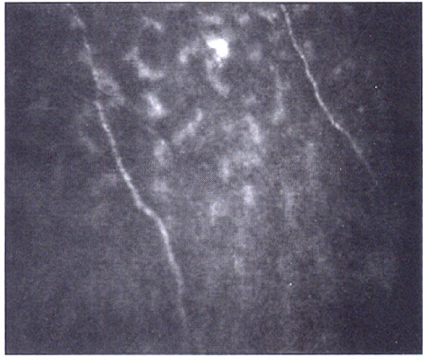
Fig. 1 Corneal subbasal nerve plexus of a patient with mild diabetic neuropathy by in vivo corneal confocal microscopy. Two nerve fibers are seen with no nerve fiber branching and mild tortuosity.
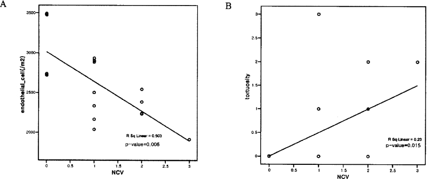
Fig. 2 Relationships between endothelial cell count (A) and tortuosity (B) and severity of neuropathy. There is a negative correlation between endothelial cell count and severity of neuropathy and a positive correlation between tortuosity and severity of neuropathy. NCV: O, normal; 1, mild neuropathy; 2, moderate neuropathy; 3, severe neuropathy.
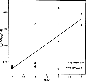
Fig. 3 Relationship between tear NGF level and neuropathy. There is a positive correlation between tear NGF level and neuropathy.
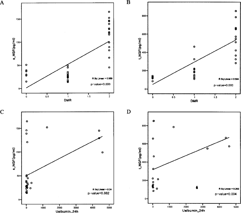
Fig. 4 Relationships between serum and tear NGF and other microvascular complications. (A, serum NGF vs. DMR; B, serum NGF vs. DMN; C, tear NGF vs. DMN; D, tear NGF vs. 24-hour albuminuria, DMR: O, normal, 1, non-proliferative retinopathy; 2, proliferative retinopathy)
Reference
-
1. Larson PR, Kronenberg HM, Melmed S, Polonsky KS. Williams Textbook of Endocrinology. 2003. 10th ed. Philadelphia: W.B. Saunders;1540–1555.2. Sugimoto K, Murakawa Y, Sima AAF. Diabetic Neuropathy: A continuing enigma. Diabetes Metab Res Rev. 2000. 16:408–433.3. Cornblath DR. Diabetic Neuropathy: Diagnostic methods. Adv Stud Med. 2004. 4:S650–S661.4. Pittenger GL, Ray M, Burcus NI, McNulty P, Basta B, Vinik AI. Intraepidermal nerve fibers are indicators of small-fiber neuropathy in both diabetic and nondiabetic patients. Diabetes Care. 2004. 27:1974–1979.5. Malik RA, Kallinikos P, Abbott CA, van Schie C, Morgan P, Efron N, Boulton AJ. Corneal Confocal Microscopy: A non-invasive surrogate of nerve fiber damage and repair in diabetic patients. Diabetologia. 2003. 46:683–688.6. Hossain P, Sachdev A, Malik RA. Early detection of diabetic peripheral neuropathy with corneal confocal microscopy. Lancet. 2005. 366:1340–1343.7. Oliveira-Soto L, Efron N. Morphology of corneal nerves using confocal microscopy. Cornea. 2001. 20(4):374–384.8. Muller LJ, Vrensen G, Pels L, Cardozo BN, Willkens B. Architecture of human corneal nerves. Invest Ophthalmol Vis Sci. 1997. 38:985–994.9. Rosenberg ME, Tervo T, Immonen IK, Muller LJ, Gronbagen-Riska C, Vesaluoma MH. Corneal structure and sensitivity in type 1 diabetes mellitus. Invest Ophthalmol Vis Sci. 2000. 41:2915–2921.10. Kallinikos P, Berbanu M, O'Donnell C, Boulton AJ, Efron N, Malik RA. Corneal nerve tortuosity in diabetic patients with neuropathy. Invest Ophthalmol Vis Sci. 2004. 45:418–422.11. Leinninger GM, Vincent AM, Feldman EL. The role of growth factor in diabetic peripheral neuropathy. J Peripher Nerv Syst. 2004. 9:76–83.12. Apfel SC. Neurotrophic factors in peripheral neuropathies: therapeutic implications. Brain Pathol. 1999. 9:393–413.13. Vinik AI. Diabetic neuropathy: pathogenesis and therapy. Am J Med. 1999. 107:17S–26S.14. Kurihara H, Shinohara H, Yoshino H, Takeda K, Shiba H. Neutrophins in cultured cells from periodontal tissues. J Periodontol. 2003. 74:76–84.15. Dobrowsky RT, Rouen S, Yu C. Altered neurotrophism in diabetic neuropathy: spelunking the caves of peripheral nerve. J Pharmacol Exp Ther. 2005. 313:485–491.16. Hellweg R, Hartung HD. Endogenous levels of nerve growth factor(NGF) are altered in experimental diabetes mellitus: a possible role for NGF in the pathogenesis of diabetic neuropathy. J Neurosci Res. 1990. 26:258–267.17. Alfel SC. Neurotrophic factors and diabetic peripheral neuropathy. Eur Neurol. 1994. 41:suppl 1. 27–34.18. Chilton L, Middlemas A, Gardiner N, Tomlinson DR. The p75 neurotrophin receptor appears in plasma in diabetic rats: characterization of a potential early test for neuropathy. Diabetologia. 2004. 47:1924–1930.19. Ordonez G, Gernandez A, Perez R, Sotelo J. Low contents of nerve growth factor in serum and submaxillary gland of diabetic mice. A possible etiological element of diabetic neuropathy. J Neurol Sci. 1994. 121:163–166.20. Pittenger G, Vinik A. Nerve growth factor and diabetic neuropathy. Exp Diabesity Res. 2003. 4:271–285.21. Lee HK, Lee KS, Kim HC, Lee SH, Kim EK. Nerve growth factor concentration and implications in photorefractive keratectomy vs. laser in situ keratomileusis. Am J Ophthalmol. 2005. 139:965–971.22. Heese K, Beck KF, Behrens MH, Pluss K, Fierlbeck W, Huwiler A, Muhl H, Geiger H, Otten U, Pfeilschifter J. Effects of high glucose on cytokine-induced nerve growth factor(NGF) expression in rat renal mesangial cells. Biochem Pharmacol. 2003. 65:293–301.23. You L, Kruse FE, Volcker HE. Neurotrophic factors in the human cornea. Invest Ophthalmol Vis Sci. 2000. 41:987–994.24. Dicou E, Nerrier V, Naud MC, deKozak Y. NGF involvement in ocular inflammation: secretion by rat resident retinal cells. Neuroreport. 1994. 6:26–28.25. Chang PY, Carrel H, Huang JS, Wang IK, Hou YC, Chen WL, Wang JY, Hu FR. Decreaseddensity of corneal basal epithelium and subbasal corneal nerve bundle changes in patients with diabetic retinopathy. Am J Opthalmol. 2006. 142:488–490.26. Karlsson M, Mayordomo R, Reichardt LF, Catsicas S, Karten HJ, Hallbook F. Nerve growth factor is expressed by postmitotic avian retinal horizontal cells and supports their survival during development in an autocrine mode of action. Development. 2001. 128:471–479.27. Patel SV, McLaren JW, Hodge DO, Bourne WM. Normal human keratocyte density and corneal thickness measurement by using confocal microscopy in vivo. Invest Ophthalmol Vis Sci. 2001. 42:333–339.28. Calvillo MP, McLaren JW, Hodge DO, Bourne WM. Corneal reinnervation after LASIK: prospective three-year longitudinal study. Invest Ophthalmol Vis Sci. 2004. 45:3991–3996.29. Erie JC, Patel SV, McLaren WJ, Hodge DO, Bourne WM. Keratocyte density in the human cornea after photorefractive keratectomy. Arch Ophthalmol. 2003. 121:770–776.30. Oh SJ. Clinical electromyography: nerve conduction studies. 1993. 2nd ed. Baltimore: Williams & Wilkins;37–53.31. Lee KY, Kim WJ, Kwon SH, Cho TY, Lee SH, Cheong KH, Park KD, Kim SM, Sunwoo IN. The usefulness of standardization of nerve conduction study in the diagnosis and follow-up of the demylinating polyneuropathy. J Korean Neurol Assoc. 1998. 16:510–518.32. McNamara NA, Brand RJ, Polse KA, Bourne WM. Corneal function during normal and high serum glucose levels in diabetes. Invest Ophthalmol Vis Sci. 1998. 39:3–17.33. Kitzmann A, Winter EJ, Nau CB, McLaren JW, Hodge DO, Bourne WM. Comparison of corneal endothelial cell images from a noncontact microscope and a scanning confocal microscope. Cornea. 2005. 24:980–984.34. Pach JM, Brubaker RF. Corneal endothelial changes in type 1 and type 2 diabetes mellitus. Am J Opthalmol. 1984. 98:401–410.35. Larsson LI, Bourne WM, Pach JM, Brubaker RF. Structure and function of the corneal endothelium in diabetes mellitus type 1 and type 2. Arch Ophthalmol. 1996. 114:9–14.36. Janiec S, Rzendkowski M, Bolek S. The relation between corneal autofluorescence, endothelial cell count and severity of the diabetic retinopathy. Int Opthalmol. 1995. 18:205–209.37. Anand P, Terenghi G, Warner G, Kopelman P, Williams-Chestnut RE, Sinicropi DV. The role of endogenous nerve growth factor in human diabetic neuropathy. Nat Med. 1996. 2:703–707.38. Diemel LT, Cai F, Anand P, Warner G, Son DR. Increased nerve growth factor mRNA in lateral calf skin biopsies from diabetic patients. Diabet Med. 1999. 16:113–118.39. Lee DA, Gross L, Wittrock DA, Windebank AJ. Localization and expression of ciliary neurotrophic factor(CNTF) in postmortem sciatic nerve from patients with motor neuron disease and diabetic neuropathy. J Neuropathol Exp Neurol. 1996. 55:915–923.40. Terenghi G, Mann D, Kopelman PG, Anand P. trkA and trkC expression is increased in human diabetic skin. Neurosci Lett. 1997. 228:33–36.41. Faradji V, Sotelo J. Low serum levels of nerve growth factor in diabetic neuropathy. Acta Neurol Scand. 1990. 81:402–406.42. Steiner P, Pfeilschifter J, Boeckh C, Radeke H, Otten U. Interleukin-1β and tumor necrosis factor-α synergistically stimulate nerve growth factor synthesis in rat mesangial cells. Am J Physiol. 1991. 261:F792–F798.43. Alpers CE, Hudkins KL, Ferguson M, Johnson RJ, Schatteman GC, Bothwell M. Nerve growth factor receptor expression in fetal, mature, and diseased human kidneys. Lab invest. 1993. 69:703–713.44. Pittenger G, Vnik A. Nerve growth factor and diabetic neuropathy. Exp Diab Res. 2003. 4:271–285.
- Full Text Links
-
- Actions
-
Cited
- CITED
-
- Close
- Share
-
- Similar articles
-
- Clinical Evaluation for Diabetic Neuropathy
- In Vivo Confocal Microscopic Findings of Corneal Tissue in Amiodarone-Induced Vortex Keratopathy
- Confocal Microscopic Findings in Posterior Polymorphous Corneal Dystrophy
- Confocal Microscopic Corneal Findings in the Normal Rabbit and Human
- The Change of Corneal Sensitivity and Recovery of Corneal Nerve after Cataract Surgery
