J Korean Ophthalmol Soc.
2014 May;55(5):770-774. 10.3341/jkos.2014.55.5.770.
A Case of Intravitreal Bevacizumab Injection for the Treatment of Choroidal Neovascularization in Morning Glory Syndrome
- Affiliations
-
- 1Department of Ophthalmology, Busan Paik Hospital, Inje University College of Medicine, Busan, Korea. ihyun@inje.ac.kr
- KMID: 2218102
- DOI: http://doi.org/10.3341/jkos.2014.55.5.770
Abstract
- PURPOSE
We report a case of intravitreal bevacizumab injection for the treatment of choroidal neovascularization in morning glory syndrome.
CASE SUMMARY
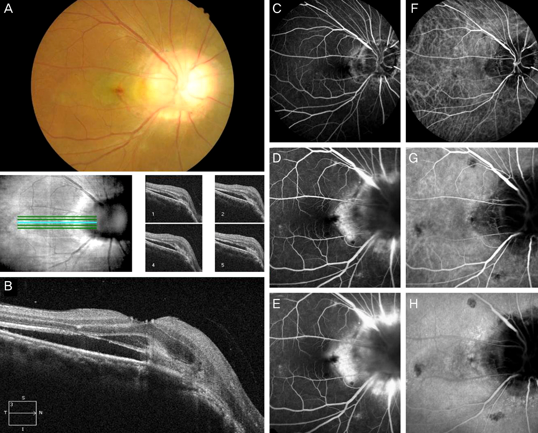
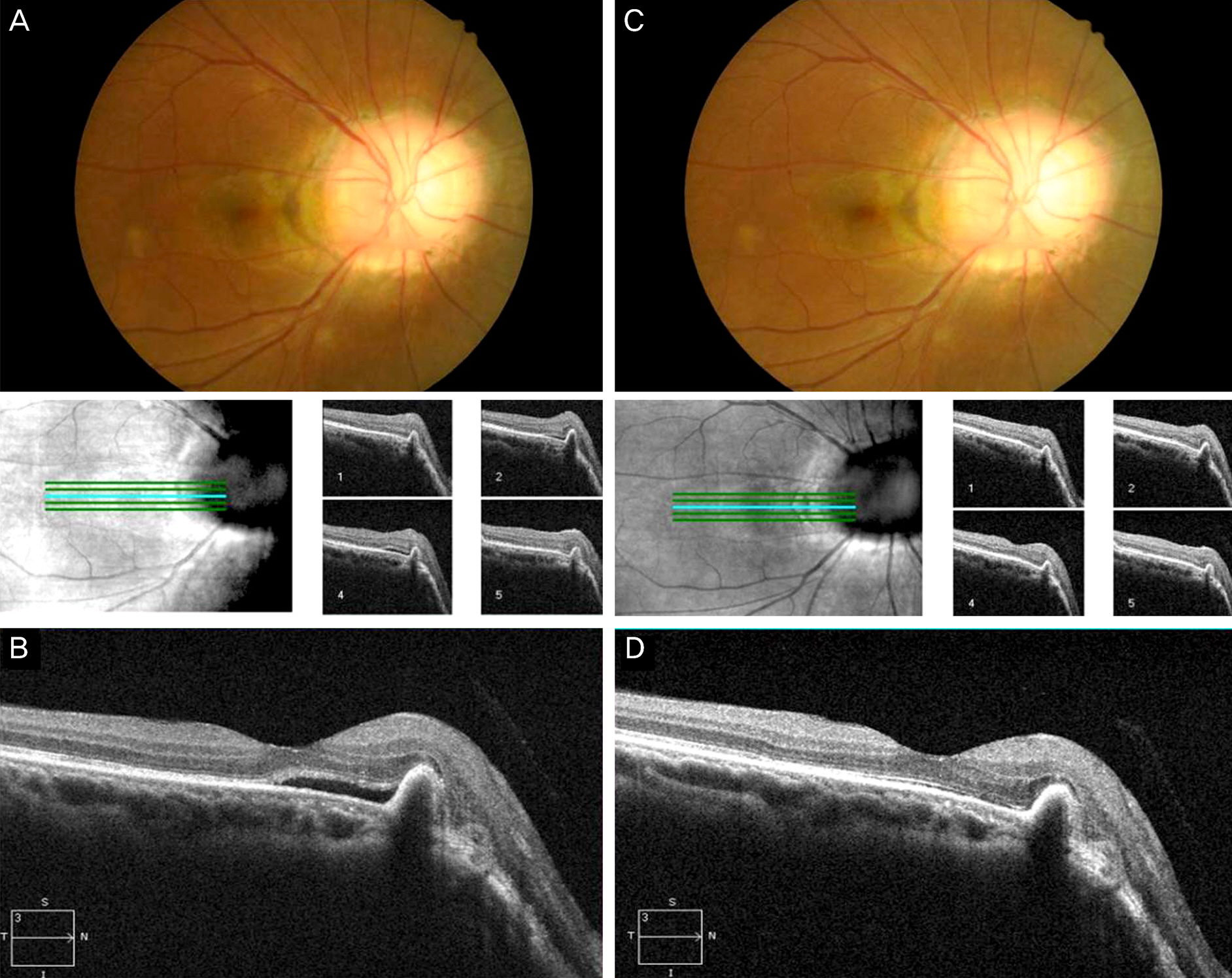
A 51-year-old male visited our hospital for a 1.5-year visual disturbance in his right eye. The patient's best-corrected visual acuity was 0.1 in the right eye. After fundus examination, we found characteristic findings of morning glory syndrome with submacular hemorrhage and serous retinal detachment in the right eye. Optical coherence tomography, fluorescein angiography and indocyanine green angiography were performed for evaluation. Retinoschisis, subretinal fluid, and choroidal neovascularization were detected, and thus bevacizumab was injected in the right eye. After intravitreal bevacizumab injection, retinoschisis was improved, and subretinal fluid was decreased. However, retinal pigment epithelial detachment was newly detected, and serous retinal detachment persisted. After 2 months, a second bevacizumab injection was performed. After these intravitreal bevacizumab injections at 1 and 2 months, visual acuity was 0.4 and 0.6, respectively. Visual acuity improved to 1.0 after 3 months. Visual acuity was maintained for at least 6 months with no relapse of choroidal neovascularization.
CONCLUSIONS
The choroidal neovascularization in morning glory syndrome was effectively treated with intravitreal bevacizumab injections.
MeSH Terms
Figure
Reference
-
References
1. Ryan SJ. Retina. 5th ed.Philadelphia: Elsevier/Mosby;2013. p. 1636–7.2. Lit ES, D'Amico DJ. Retinal manifestations of morning glory disc syndrome. Int Ophthalmol Clin. 2001; 41:131–8.
Article3. Chang S, Gregory-Roberts E, Chen R. Retinal detachment associated with optic disc colobomas and morning glory syndrome. Eye (Lond). 2012; 26:494–500.
Article4. Chang S, Haik BG, Ellsworth RM, et al. Treatment of total retinal detachment in morning glory syndrome. Am J Ophthalmol. 1984; 97:596–600.
Article5. Choudhry N, Ramasubramanian A, Shields CL, et al. Spontaneous resolution of retinal detachment in morning glory disk anomaly. J AAPOS. 2009; 13:499–500.6. Campos CC, Manzanaro PG, Rodilla JY, Martin Rodrigo JC. Retinal detachment associated with morning glory syndrome. Arch Soc Esp Oftalmol. 2011; 86:295–9.7. Akamine T, Doi M, Takahashi H, et al. Morning glory syndrome with peripheral exudative retinal detachment. Retina. 1997; 17:73–4.8. Sobol WM, Bratton AR, Rivers MB, Weingeist TA. Morning glory disk syndrome associated with subretinal neovascular membrane formation. Am J Ophthalmol. 1990; 110:93–4.
Article9. Chuman H, Nao-i N, Sawada A. A case of morning glory syndrome associated with contractile movement of the optic disc and subretinal neovascularization. Nippon Ganka Gakkai Zasshi Sep. 1996; 100:705–9.10. Browning DJ, Fraser CM. Ocular conditions associated with peripapillary subretinal neovascularization, their relative frequencies, and associated outcomes. Ophthalmology. 2005; 112:1054–61.
Article
- Full Text Links
- Actions
-
Cited
- CITED
-
- Close
- Share
- Similar articles
-
- Effect of High-dose Intravitreal Bevacizumab Injection on Refractory Idiopathic Choroidal Neovasculariz
- Choroidal Neovascularization in a Patient with Best Disease
- Long-term Therapeutic Effect of Intravitreal Bevacizumab (Avastin) on Myopic Choroidal Neovascularization
- Intravitreal Bevacizumab Injection for the Treatment of Choroidal Neovascularization Secondary to Candida Chorioretinitis
- Multifocal Electroretinogram Findings after Intravitreal Bevacizumab Injection in Choroidal Neovascularization of Age-Related Macular Degeneration


