J Korean Ophthalmol Soc.
2009 Jun;50(6):919-922. 10.3341/jkos.2009.50.6.919.
Effect of Nitric Oxide on the Survival of R28 Cells
- Affiliations
-
- 1Department of Ophthalmology, Catholic University of Daegu College of Medicine, Daegu, Korea. jwkim@cu.ac.kr
- KMID: 2212371
- DOI: http://doi.org/10.3341/jkos.2009.50.6.919
Abstract
-
PURPOSE: To evaluate the effect of nitric oxide (NO) on the survival of R28 cells.
METHODS
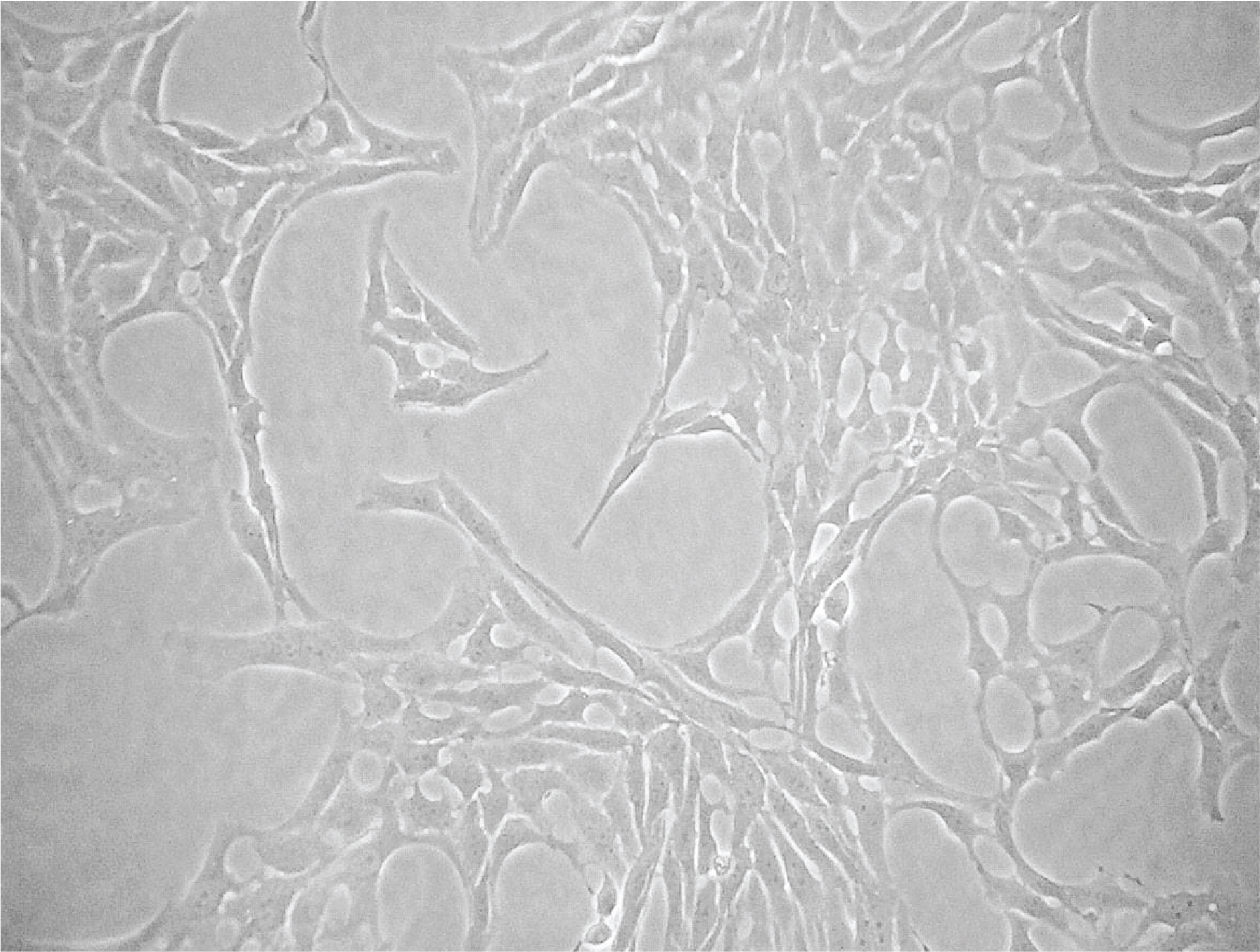
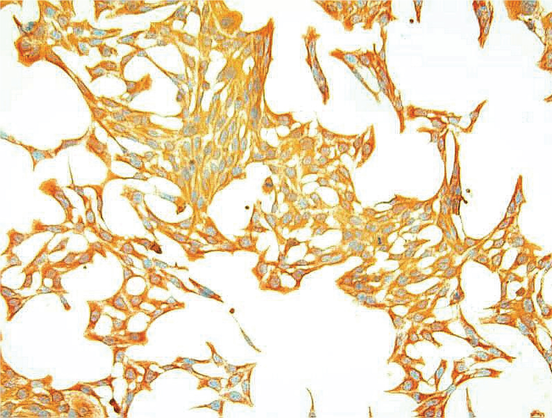
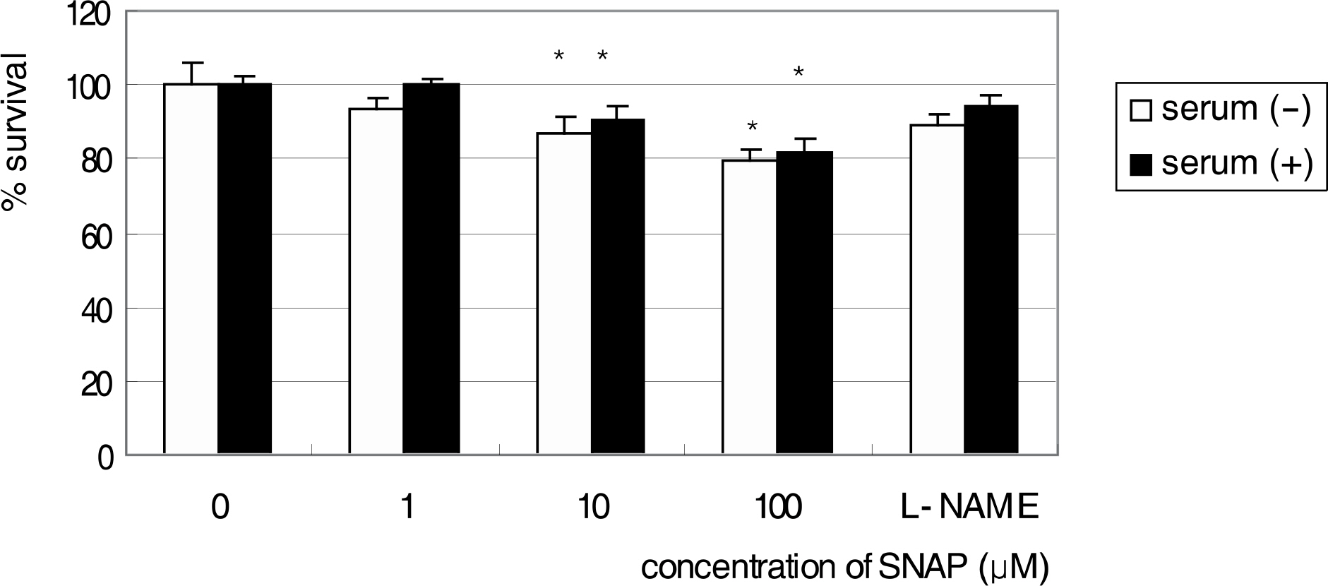
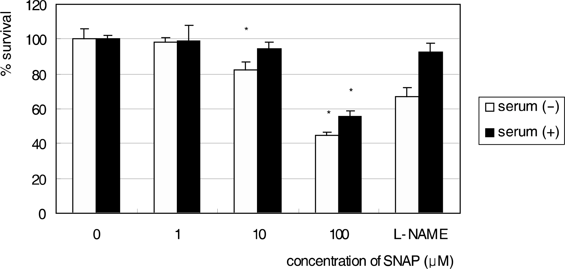
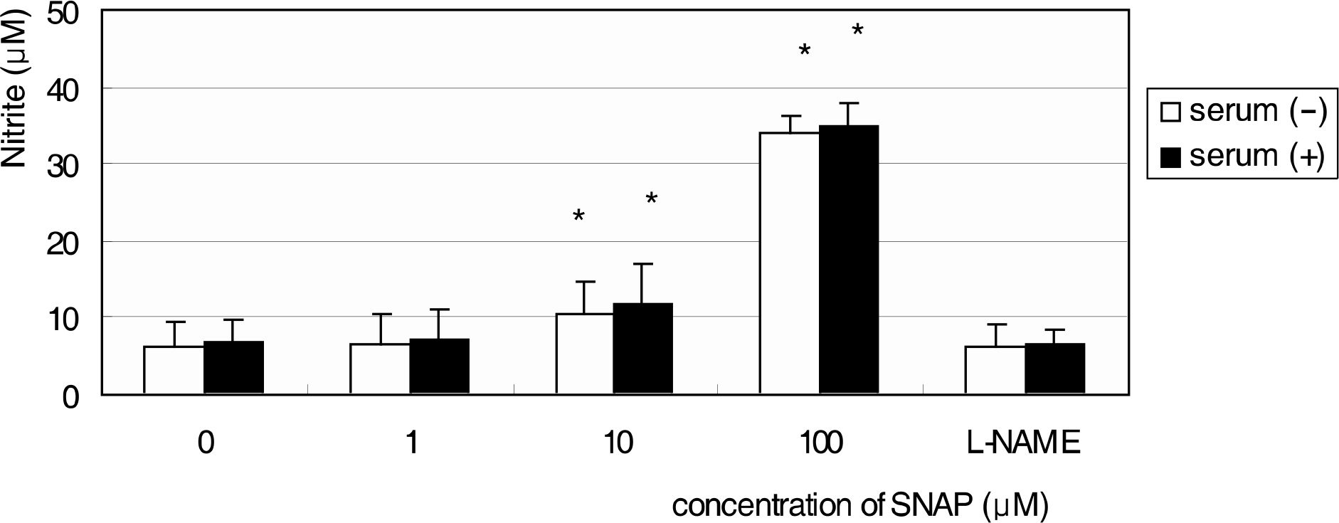
After immunostaining for GFAP, vimentin, S-100, and neurofilament, R28 cells were exposed to S-nitroso-N-acetyl-D, L-penicillamine (SNAP) at various concentrations, with and without the NO inhibitor, Nomega-Nitro-L-arginine methyl ester (L-NAME) for 1 and 3 days. Cellular survival of R28 cells and the production of NO were quantified by rapid colorimetric assays using the MTT and Griess assay, respectively. To evaluate the effect of serum, 10% serum or serum-free media were used separately.
RESULTS
R28 cells showed strong immunoreactivity to GFAP and vimentin compared to S-100 or neurofilament. SNAP inhibited the survival of R28 cells in a dose-dependent manner, and this effect of NO on the cellular survival was abolished by L-NAME. These results were similar after exposure for 1 and 3 days, regardless of the presence of serum in the media.
CONCLUSIONS
The current results suggest that NO decreased the survival of R28 cells. Further studies are necessary to evaluate the mechanism of cytotoxicity of the R28 cells.
Keyword
MeSH Terms
Figure
Reference
-
References
1. Moncada S, Palmer RM, Higgs EA. Nitric oxide: physiology, pathophysiology, and pharmacology. Pharmacol Rev. 1991; 43:109–42.2. Bredt DS, Snyder SH. Nitric oxide: a physiologic messenger molecule. Annu Rev Biochem. 1994; 63:175–95.
Article3. Brüne B, Knethen A, Sandau KB. Nitric oxide and its role in apoptosis. Eur J Pharmacol. 1998; 351:261–72.
Article4. Beck KF, Eberhardt W, Frank S, et al. Inducible NO synthase: Role in cell signaling. J Exp Biol. 1999; 202:645–53.5. Becquet F, Courtois Y, Goureau O. Nitric oxide in the eye: multifaceted roles and diverse outcomes. Surv Ophthalmol. 1997; 42:71–82.
Article6. Meyer P, Champion C, Schlotzer-Schrehardt U, et al. Localization of nitric oxide synthase isoforms in porcine ocular tissues. Curr Eye Res. 1999; 18:375–80.
Article7. Geyer O, Podos SM, Mittag T. Nitric oxide synthase activity in tissues of the bovine eyes. Graefes Arch Clin Exp Ophthalmol. 1997; 235:786–93.8. Nathanson JA, McKee M. Identification of an extensive system of nitric oxide-producing cells in the ciliary muscle and outflow pathway. Invest Ophthalmol Vis Sci. 1995; 36:1765–73.9. Quigley HA, Nickells RW, Kerrigan LA, et al. Retinal ganglion cell death in experimental glaucoma and after axotomy occurs by apoptosis. Invest Ophthalmol Vis Sci. 1995; 36:774–86.10. Otori Y, Wei J-Y, Barnstable CJ. Neurotoxic effects of low doses of glutamate on purified rat retinal ganglioncells. Invest Ophthalmol Vis Sci. 1998; 39:972–81.11. Morgan J, Caprioli J, Koseki Y. Nitric oxide mediates excitotoxic and anoxic damage in rat retinal ganglion cells cocultured with astroglia. Arch Ophthalmol. 1999; 117:1524–9.
Article12. Vorwerk CK, Hyman BT, Miller JW, et al. The role of neuronal and endothelial nitric oxide synthase in retinal excitotoxicity. Invest Ophthalmol Vis Sci. 1997; 38:2038–44.13. Seigel GM. The golden age of retinal cell culture. Mol Vis. 1999; 5:4.14. Seigel GM, Mutchler AL, Imperato EL. Expression of glial markers in retinal precusor cell line. Mol Vis. 1996; 2:2.15. Green LC, Wagner DA, Glogoski J, et al. Analysis of nitrate, nitrite and [15N]nitrate in biologic fluids. Anal Biochem. 1982; 126:131–8.16. Nathanson JA, McKee M. Alterations of ocular nitric oxide synthase in human glaucoma. Invest Ophthalmol Vis Sci. 1995; 36:1774–84.17. Neufeld AH, Hernandez MR, Gonzalez M. Nitric oxide synthase in the human glaucomatous optic nerve head. Arch Ophthalmol. 1997; 115:497–503.
Article18. Hu DN, Ritch R. Tissue culture of adult human retinal ganglion cells. J Glaucoma. 1997; 6:37–43.
Article19. Barres BA, Silverstein BE, Corey DP, Chun LL. Immunological, morphological, and electrophysiological variation among retinal ganglion cells purified by panning. Neuron. 1988; 1:791–803.
Article20. Seigel GM, Takahashi M, Adamus G, McDaniel T. Intraocular transplantation of E1A-immortalized retinal precusor cells. Cell Transplant. 1998; 7:559–66.21. Seigel GM, Chiu L, Paxhia A. Inhibition of neuroretinal cell death by insulinlike growth factor-1 and its analogs. Mol Vis. 2000; 6:157–63.22. Barber AJ, Nakamura M, Wolpert EB, et al. Insulin rescues retinal neurons from apoptosis by a phosphatidyl 3-kinase/Akt-mediated mechanism that reduces the activation of Caspases-3. J Biol Chem. 2001; 276:32814–21.23. Nakamura M, Barber AJ, Antonetti DA, et al. Excessive hexo-samines block the neuroprotective effect of insulin and induce apoptosis in retinal neurons. J Biol Chem. 2001; 276:43748–55.
Article24. Sun W, Seigel GM, Salvi RJ. Retinal precusor cells express functional ionotropic glutamate and GABA receptors. Neuroreport. 2002; 13:2421–4.25. Kawasaki A, Otori Y, Barnstable CJ. Müller cell protection of rat retinal ganglion cells from glutamate and nitric oxide neurotoxicity. Invest Ophthalmol Vis Sci. 2000; 41:3444–50.
- Full Text Links
- Actions
-
Cited
- CITED
-
- Close
- Share
- Similar articles
-
- Effect of High Glucose on the Production of Reactive Oxygen Species in R28 Cells
- Effect of Erythropoietin on the Production of Nitric Oxide in Trabecular Meshwork Cells
- Effect of Genistein on the Survival and Production of Nitric Oxide in Trabecular Meshwork Cells
- Expression of Constitutive Nitric Oxide Synthase by Gastrointestinal Epithelial Cells
- Effect of beta-adrenergics on the Survival and Production of Nitric Oxide in the Cultured Trabecular Meshwork Cells

