Investigation of bone formation using calcium phosphate glass cement in beagle dogs
- Affiliations
-
- 1Department of Periodontology, Research Institute for Periodontal Regeneration, Yonsei University College of Dentistry, Seoul, Korea. shchoi726@yuhs.ac
- 2Department and Research Institute of Dental Biomaterials and Bioengineering, Yonsei University College of Dentistry, Seoul, Korea.
- KMID: 2212139
- DOI: http://doi.org/10.5051/jpis.2010.40.3.125
Abstract
- PURPOSE
Among available biomaterials, bioceramics have drawn special interest due to their bioactivity and the possibility of tailoring their composition. The degradation rate and formulation of bioceramics can be altered to mimic the compositions of the mineral phase of bone. The aim of this study was to investigate the bone formation effect of amorphous calcium phosphate glass cement (CPGC) synthesized by a melting and quenching process.
METHODS
In five male beagle dogs, 4 x 4 mm 1-wall intrabony defects were created bilaterally at the mesial or distal aspect of the mandibular second and fourth premolars. Each of the four defects was divided according to graft materials: CPGC with collagen membrane (CM), biphasic calcium phosphate (BCP) with CM, CM alone, or a surgical flap operation only. The dogs were sacrificed 8 weeks post-surgery, and block sections of the defects were collected for histologic and histometric analysis.
RESULTS
There were significant differences in bone formation and cementum regeneration between the experimental and control groups. In particular, the CPGC and BCP groups showed greater bone formation than the CM and control groups.
CONCLUSIONS
In conclusion, CPGC was replaced rapidly with an abundant volume of new bone; CPGC also contributed slightly to regeneration of the periodontal apparatus.
Keyword
MeSH Terms
-
Animals
Bicuspid
Biocompatible Materials
Bone Substitutes
Calcium
Calcium Phosphates
Collagen
Dental Cementum
Dogs
Freezing
Glass
Humans
Hydrazines
Hydroxyapatites
Male
Membranes
Osteogenesis
Regeneration
Surgical Flaps
Transplants
Biocompatible Materials
Bone Substitutes
Calcium
Calcium Phosphates
Collagen
Hydrazines
Hydroxyapatites
Figure
-
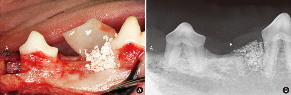
Figure 1 Clinical photograph (A) and radiograph (B) showing surgically prepared 1-wall intrabony defects at the mesial aspect of the second premolar and fourth premolar (A: collagen membrane group or control group, B: calcium phosphate glass cement group or biphasic calcium phosphate).
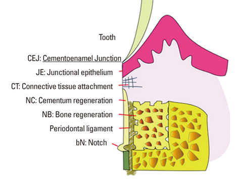
Figure 2 Schematic diagram depicting the landmarks and the parameters used in histometric analysis. The heights of new bone, new cementum, epithelial, and connective tissue attachment in 4 × 4 mm 1-wall intrabony defects were measured using an automated image analysis system.

Figure 3 Surgical sections from the calcium phosphate glass cement (CPGC) group. (A) Histologic view of the CPGC group. Most particles were resorbed and new bone was formed above the notch (H&E, ×20; base of reference notch [bN]: arrow; height of new bone: black diamond; CPGC particle: black star; bar = 2 mm). (B) Histologic view of magnified black square area (×200). Osteoblast-like cells were observed around remaining particles. Peripheral new bone was woven bone with isolated osteocytes (H&E, ×200; CPGC particle: black star; NB: new bone; WB: woven bone; bar = 0.1 mm).
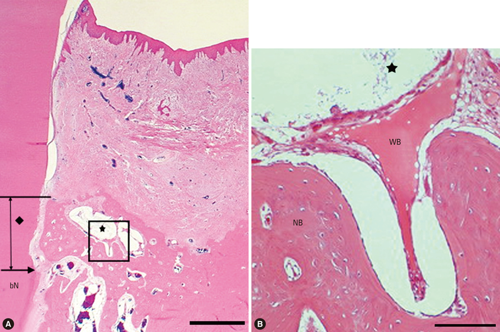
Figure 4 Surgical sections from the biphasic calcium phosphate (BCP) group. (A) Histologic view of BCP group. There were more remaining particles than in the calcium phosphate glass cement (CPGC) group. New bone was formed above the notch (H&E, ×20; base of reference notch [bN]: arrow; the height of new bone: black diamond; BCP particle: black star; bar = 2 mm). (B) Histologic view of magnified black square area (×200). Multi-nucleated giant cells were arranged around particles and woven bone was formed (H&E, ×200; BCP particle: black star; NB: new bone; WB: woven bone; bar = 0.1 mm).
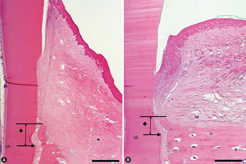
Figure 5 Surgical sections from the collagen membrane (CM) group and the control group. (A) Histologic view of the CM group. A small amount of new bone was formed above the notch. Thick connective tissue was present (H&E, ×20; base of reference notch [bN]: arrow; height of new bone: black diamond; bar = 2 mm). (B) Histologic view of the control group. There was very little new bone above the notch. Long junctional epithelium was observed and inflammatory cell infiltrate was relatively strong (H&E, ×20; base of reference notch [bN]: arrow; height of new bone: black diamond; bar = 2 mm).
Reference
-
1. Parikh SN. Bone graft substitutes in modern orthopedics. Orthopedics. 2002. 25:1301–1309.
Article2. Ahlmann E, Patzakis M, Roidis N, Shepherd L, Holtom P. Comparison of anterior and posterior iliac crest bone grafts in terms of harvest-site morbidity and functional outcomes. J Bone Joint Surg Am. 2002. 84A:716–720.
Article3. Kurashina K, Kurita H, Wu Q, Ohtsuka A, Kobayashi H. Ectopic osteogenesis with biphasic ceramics of hydroxyapatite and tricalcium phosphate in rabbits. Biomaterials. 2002. 23:407–412.
Article4. Jarcho M, Kay JF, Gumaer KI, Doremus RH, Drobeck HP. Tissue, cellular and subcellular events at a bone-ceramic hydroxylapatite interface. J Bioeng. 1977. 1:79–92.5. Rabalais ML Jr, Yukna RA, Mayer ET. Evaluation of durapatite ceramic as an alloplastic implant in periodontal osseous defects. I. Initial six-month results. J Periodontol. 1981. 52:680–689.
Article6. Gao H, Tan T, Wang D. Effect of composition on the release kinetics of phosphate controlled release glasses in aqueous medium. J Control Release. 2004. 96:21–28.
Article7. Bagambisa FB, Joos U, Schilli W. Interaction of osteogenic cells with hydroxylapatite implant materials in vitro and in vivo. Int J Oral Maxillofac Implants. 1990. 5:217–226.8. Kim YK, Yun PY, Lim SC, Kim SG, Lee HJ, Ong JL. Clinical evaluations of OSTEON as a new alloplastic material in sinus bone grafting and its effect on bone healing. J Biomed Mater Res B Appl Biomater. 2008. 86:270–277.
Article9. Hosono H, Abe Y. Porous glass-ceramics composed of a titanium phosphate crystal skeleton: a review. J Non-Cryst Solids. 1995. 190:185–197.
Article10. Lee BH, Kim MC, Kim KN, LeGeros RZ, Lee YK. Biodegradable bone cement using calcium phosphate glass. Key Eng Mater. 2006. 309-311:861–864.
Article11. Nery EB, Eslami A, Van Swol RL. Biphasic calcium phosphate ceramic combined with fibrillar collagen with and without citric acid conditioning in the treatment of periodontal osseous defects. J Periodontol. 1990. 61:166–172.
Article12. Bohner M. Physical and chemical aspects of calcium phosphates used in spinal surgery. Eur Spine J. 2001. 10:Suppl 2. S114–S121.
Article13. Bunker BC, Arnold GW, Wilder JA. Phosphate glass dissolution in aqueous solutions. J Non-Cryst Solids. 1984. 64:291–316.
Article14. Murphy KG, Gunsolley JC. Guided tissue regeneration for the treatment of periodontal intrabony and furcation defects. A systematic review. Ann Periodontol. 2003. 8:266–302.
Article15. Dias AG, Lopes MA, Gibson JR, Santos JD. In vitro degradation studies of calcium phosphate glass ceramics prepared by controlled crystallization. J Non-Cryst Solids. 2003. 330:81–89.
Article16. Maus U, Andereya S, Gravius S, Ohnsorge JA, Niedhart C, Siebert CH. BMP-2 incorporated in a tricalcium phosphate bone substitute enhances bone remodeling in sheep. J Biomater Appl. 2008. 22:559–576.
Article17. Wolff KD, Swaid S, Nolte D, Bockmann RA, Holzle F, Muller-Mai C. Degradable injectable bone cement in maxillofacial surgery: indications and clinical experience in 27 patients. J Craniomaxillofac Surg. 2004. 32:71–79.
Article
- Full Text Links
-
- Actions
-
Cited
- CITED
-
- Close
- Share
-
- Similar articles
-
- Effects on the Tissue Reaction Using GI Cement in the Maxillary Grade II Furcation in the Beagle Dogs
- Novel Calcium Phosphate Glass for Hard-Tissue Regeneration
- The Biological Effects of Calcium Phosphate Coated Implant for Osseointegration in Beagle Dogs
- Histologic findings of three-wall intrabony defects around dental implants using different grafting materials in beagle dogs
- The effects of noncrystalline calcium phosphate glass on the healing of 1-wall intrabony defects in beagle dogs
