J Korean Soc Magn Reson Med.
2013 Sep;17(3):207-214. 10.13104/jksmrm.2013.17.3.207.
Magnetic Resonance Imaging of Breast Cancer Patients with BRCA Mutation
- Affiliations
-
- 1Department of Radiology and Research Institute of Radiology, Asan Medical Center, College of Medicine, University of Ulsan, Seoul, Korea. jhcha@amc.seoul.kr
- 2Department of General Surgery, Asan Medical Center, College of Medicine, University of Ulsan, Seoul, Korea.
- 3Department of Internal Medicine, Asan Medical Center, College of Medicine, University of Ulsan, Seoul, Korea.
- KMID: 2206919
- DOI: http://doi.org/10.13104/jksmrm.2013.17.3.207
Abstract
- PURPOSE
To evaluate the MRI findings of breast cancer with BRCA mutation.
MATERIALS AND METHODS
We collected information of the breast cancer patients who underwent the test for BRCA gene mutation as well as preoperative breast MRI from January 2007 to December 2010. A total of 185 patients were enrolled; 33 of these patients had BRCA mutations and 152 patients did not. Among them, a total of 231 breast cancers were detected. Images of the 47 breast cancers with BRCA mutation and of the 184 breast cancers without mutations were evaluated to compare the morphologic and enhancement features on MRI.
RESULTS
With MR imaging, there were no significant difference in morphologic characteristic between two groups. However, enhancement pattern in the group with BRCA mutation were more likely to have persistent enhancement (p < 0.233), and LN metastasis was more common in breast cancers without BRCA mutation. Breast cancers with BRCA 2 mutation tend to show more persistent enhancement pattern than BRCA 1 mutation.
CONCLUSION
In breast cancer patients with BRCA mutation, MRI didn't show significant difference in morphologic characteristics, however breast cancers with BRCA gene mutation carriers tend to have benign morphologic features on MRI, such as Type 1 kinetic curve enhancement.
MeSH Terms
Figure
-
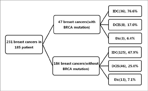
Fig. 1 Dominant Pathologic Types for Tumors.

Fig. 2 a. An axial subtraction MR image of a 32-year-old BRCA 2 gene mutation carrier. In the right breast outer portion, an oval shaped, homogeneous enhancing lesion is visible with longest diameter of 8 mm. b. The kinetic analysis reveals delayed persistent enhancement of the lesion.
Reference
-
1. Choi DH, Lee MH, Bale AE, Carter D, Haffty BG. Incidence of BRCA1 and BRCA2 mutations in young Korean breast cancer patients. J Clin Oncol. 2004; 22:1638–1645.2. Stratton MR. Pathology of familial breast cancer: differences between breast cancers in carriers of BRCA1 or BRCA2 mutations and sporadic cases. Breast cancer linkage consortium. Lancet. 1997; 349:1505–1510.3. Feig SA, D'Orsi CJ, Hendrick RE, et al. American college of radiology guidelines for breast cancer screening. AJR Am J Roentgenol. 1998; 171:29–33.4. Burke W, Daly M, Garber J, et al. Recommendations for follow-up care of individuals with an inherited predisposition to cancer. Ii. BRCA1 and BRCA2. Cancer genetics studies consortium. JAMA. 1997; 277:997–1003.5. Kriege M, Brekelmans CT, Boetes C, et al. Efficacy of MRI and mammography for breast-cancer screening in women with a familial or genetic predisposition. N Engl J Med. 2004; 351:427–437.6. Kriege M, Brekelmans CT, Boetes C, et al. MRI screening for breast cancer in women with familial or genetic predisposition: design of the Dutch National Study (MRISC). Fam Cancer. 2001; 1:163–168.7. Plevritis SK, Kurian AW, Sigal BM, et al. Cost-effectiveness of screening BRCA1/2 mutation carriers with breast magnetic resonance imaging. JAMA. 2006; 295:2374–2384.8. Warner E, Plewes DB, Shumak RS, et al. Comparison of breast magnetic resonance imaging, mammography, and ultrasound for surveillance of women at high risk for hereditary breast cancer. J Clin Oncol. 2001; 19:3524–3531.9. Kuhl CK, Schmutzler RK, Leutner CC, et al. Breast MR imaging screening in 192 women proved or suspected to be carriers of a breast cancer susceptibility gene: preliminary results. Radiology. 2000; 215:267–279.10. Saslow D, Boetes C, Burke W, et al. American cancer society guidelines for breast screening with MRI as an adjunct to mammography. CA Cancer J Clin. 2007; 57:75–89.11. Warner E, Plewes DB, Hill KA, et al. Surveillance of BRCA1 and BRCA2 mutation carriers with magnetic resonance imaging, ultrasound, mammography, and clinical breast examination. JAMA. 2004; 292:1317–1325.12. Veltman J, Mann R, Kok T, et al. Breast tumor characteristics of BRCA1 and BRCA2 gene mutation carriers on MRI. Eur Radiol. 2008; 18:931–938.13. Schrading S, Kuhl CK. Mammographic, US, and MR imaging phenotypes of familial breast cancer. Radiology. 2008; 246:58–70.14. Blaichman J, Marcus JC, Alsaadi T, El-Khoury M, Meterissian S, Mesurolle B. Sonographic appearance of invasive ductal carcinoma of the breast according to histologic grade. AJR Am J Roentgenol. 2012; 199:W402–W408.15. Gilbert FJ, Warren RM, Kwan-Lim G, et al. Cancers in BRCA1 and BRCA2 carriers and in women at high risk for breast cancer: MR imaging and mammographic features. Radiology. 2009; 252:358–368.16. Son BH, Ahn SH, Park SK, Kim SW. Hereditary breast cancer in Korea: a review of the literature. J Breast Cancer. 2008; 11:1–9.
- Full Text Links
-
- Actions
-
Cited
- CITED
-
- Close
- Share
-
- Similar articles
-
- Association between BRCA Mutation Status, Pathological Findings, and Magnetic Resonance Imaging Features in Patients with Breast Cancer at Risk for the Mutation
- Occurrence of contralateral breast cancer in a BRCA-positive breast cancer patient who underwent free TRAM flap reconstruction: a case report
- A Rare Case of Male Primary Breast Lymphoma
- Experience with Bilateral Risk-Reducing Mastectomy for an Unaffected BRCA Mutation Carrier
- Is the BRCA Germline Mutation a Prognostic Factor in Korean Patients with Early-onset Breast Carcinomas?
