A Rare Case of Male Primary Breast Lymphoma
- Affiliations
-
- 1Department of Radiodiagnosis, Government Medical College and Hospital, Nagpur, India. drjawahar_12@rediffmail.com
- KMID: 2175735
- DOI: http://doi.org/10.4048/jbc.2011.14.4.333
Abstract
- Primary lymphoma of the breast is a rare occurrence because of the paucity of lymphoid tissue in the breast and is an even rarer entity in the male breast. Imaging, along with tissue diagnosis goes a long way in diagnosing breast lymphoma which has a significantly different management scheme than other breast neoplasms with respect to radio-chemotherapy rather than surgical resection. We present a case of primary male breast lymphoma which was evaluated with magnetic resonance imaging as well as other conventional imaging modalities and was treated by chemotherapy with a 7-month follow-up.
Keyword
MeSH Terms
Figure
-
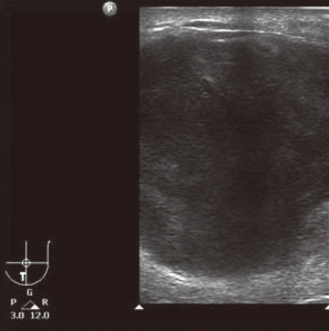
Figure 1 High frequency ultrasound using 12 to 3 MHz broadband linear array transducer revealed a well-defined lobulated relatively homogenous, hypoechoic mass.

Figure 2 On color Doppler, the lesion showed mild vascularity.
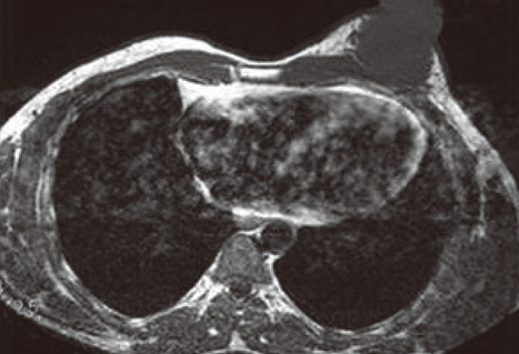
Figure 3 T1-weighted MR image showing a well-defined isointense, lobulated lesion and maintained fat planes with the underlying pectoralis muscle.
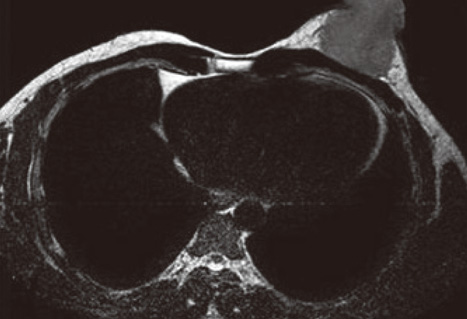
Figure 4 T2-weighted MR image showing a mildly hyperintense lesion compared to muscle.
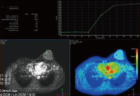
Figure 5 Dynamic MR imaging showing a Type II perfusion curve.

Figure 6 H&E stained image showing diffuse large B cell lymphoma (H&E, ×100).

Figure 7 Immunohistochemical study showing diffuse positivity of diffuse round cells for CD 20 (Immunohistochemical stain, ×600).
Cited by 1 articles
-
Diffuse Large B-Cell Lymphoma with Involvement of the Breast and Testis in a Male Patient
Eunji Choi, Jae-Cheol Jo, Dok Hyun Yoon, Shin Kim, Kyoungmin Lee, Jooryung Huh, Chan-Sik Park, Sang Wook Lee, Cheolwon Suh
Cancer Res Treat. 2015;47(3):539-543. doi: 10.4143/crt.2013.245.
Reference
-
1. Issacson PG, Norton AJ. Issacson PG, Norton AJ, editors. Lymphoma of the breast. Extranodal Lymphomas. 1994. Edinburgh: Churchill-Livingstone;289–298.2. Stavros AT. Breast Ultrasound. 2004. Philadelphia: Lippincott Williams & Wilkins;864.3. Liberman L, Giess CS, Dershaw DD, Louie DC, Deutch BM. Non-Hodgkin lymphoma of the breast: imaging characteristics and correlation with histopathologic findings. Radiology. 1994. 192:157–160.
Article4. Domchek SM, Hecht JL, Fleming MD, Pinkus GS, Canellos GP. Lymphomas of the breast: primary and secondary involvement. Cancer. 2002. 94:6–13.5. Wiseman C, Liao KT. Primary lymphoma of the breast. Cancer. 1972. 29:1705–1712.
Article6. Yang WT, Lane DL, Le-Petross HT, Abruzzo LV, Macapinlac HA. Breast lymphoma: imaging findings of 32 tumors in 27 patients. Radiology. 2007. 245:692–702.
Article7. Tozaki M, Maruyama K. H MR spectroscopy of the breast. MAGNETOM Flash. 2007. (3):41–43.8. Wu X, Kellokumpu-Lehtinen PL, Pertovaara H, Korkola P, Soimakallio S, Eskola H, et al. Diffusion-weighted MRI in early chemotherapy response evaluation of patients with diffuse large B-cell lymphoma - a pilot study: comparison with 2-deoxy-2-fluoro-D-glucose-positron emission tomography/computed tomography. NMR Biomed. Epub 2011 Mar 8. DOI: 10.1002/nbm.1689.9. Morris EA. Edelman RR, Hesselink JR, Zlatkin MB, Crues JV, editors. Breast cancer. Clinical Magnetic Resonance Imaging. 2006. 3rd ed. Philadelphia: Saunders;2440–2444.10. Mpallas G, Simatos G, Tasidou A, Patra E, Galateros G, Lakiotis G, et al. Primary breast lymphoma in a male patient. Breast. 2004. 13:436–438.
Article
- Full Text Links
-
- Actions
-
Cited
- CITED
-
- Close
- Share
-
- Similar articles
-
- Primary Mucosa-Associated Lymphoid Tissue Lymphoma of the Breast with Synchronous Contralateral Invasive Breast Cancer: A Case Report
- Primary Breast Lymphoma in an Immunocompromised Male Patient: A Case Report
- Primary Follicular Lymphoma in a Male Breast: A Case Report
- Primary breast lymphoma
- Primary Peripheral T-cell Lymphoma of the Breast: Radiologic and Pathologic Findings
