Primary Peripheral T-cell Lymphoma of the Breast: Radiologic and Pathologic Findings
- Affiliations
-
- 1Department of Radiology, Korea University College of Medicine, Seoul, Korea. krcho@korea.ac.kr
- 2Department of Pathology, Korea University College of Medicine, Seoul, Korea.
- 3Department of Surgery, Korea University College of Medicine, Seoul, Korea.
Abstract
- Primary breast lymphoma is a rare disease entity, particularly the T-cell type. There have been many case reports of primary breast lymphomas; however, these are mostly pathologic reports, with only a few reports in radiology literature. To the best of our knowledge, this is the first report on the radiologic features of primary T-cell type breast lymphoma, including mammography, ultrasonography, MR imaging, and 18 fluorodeoxyglucose positron emission tomography/computed tomography scan. The radiologic findings are rather unique for this T-cell lymphoma compared to B cell type.
Keyword
MeSH Terms
Figure
-
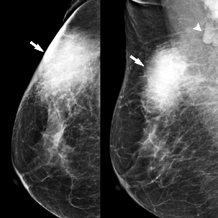
Figure 1 Mammography shows an ill-defined, irregular shaped hyperdense mass (arrow) in the right upper outer quadrant, accompanied by slight thickening of the overlying skin and ipsilateral lymphadenopathy (arrowhead).
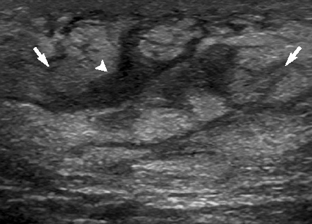
Figure 2 Ultrasonography shows an ill-defined hyperechoic lesion (arrow) with tubular shaped branching hypoechogenecities (arrowhead) without focal mass.
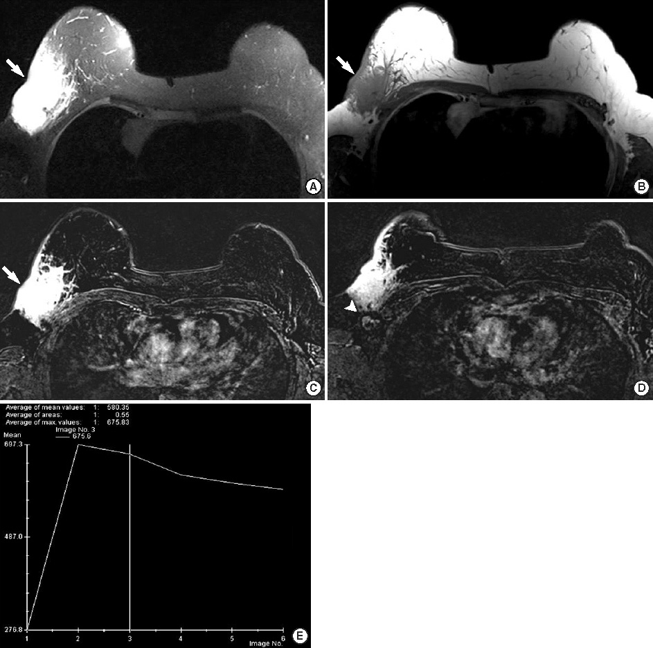
Figure 3 MRI demonstrates an ill-defined irregular mass showing very high signal intensity on T2 WI (A, arrow) and low signal intensity on T1 WI (B, arrow) in the upper outer quadrant of right breast. Early subtraction images (C, D) show a well-enhanced mass (C, arrow). An enhancing enlarged lymph node is visible in the right axilla (D, arrowhead), with well-delineated skin thickening. This shows a type III pattern time-signal intensity curve (early enhancement and delayed wash-out) on dynamic study (E).
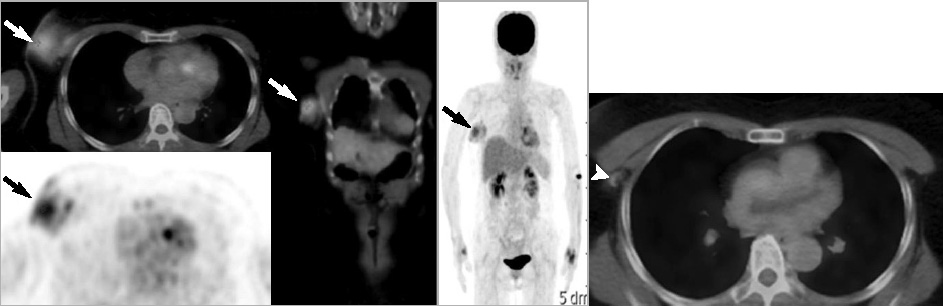
Figure 4 F-18 FDG PET/CT reveals a moderate hypermetabolic lesion (arrow) and maximum standard uptake value (SUV) ranges from 3.3 to 3.8. A focal hypermetabolic lesion measuring 2.3 of maximum SUV is also visible at the right axilla (arrowhead).
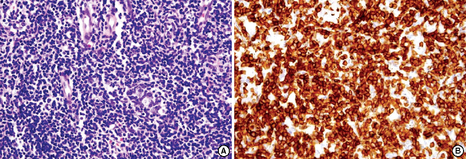
Figure 5 Histopathologic examination shows diffuse dense infiltration of small and large lymphoid cells (A, ×200), composed of predominantly CD3-positive T-cells (B, ×200).
Reference
-
1. Arber DA, Simpson JF, Weiss LM, Rappaport H. Non-Hodgkin's lymphoma involving the breast. Am J Surg Pathol. 1994. 18:288–295.
Article2. Uesato M, Miyazawa Y, Gunji Y, Ochiai T. Primary non-Hodgkin's lymphoma of the breast: report of a case with special reference to 380 cases in the Japanese literature. Breast Cancer. 2005. 12:154–158.
Article3. Petrek JA. Harris JR, editor. Lymphoma. Breast Diseases. 1991. 2nd ed. Philadelphia: Lippincott;806–807.4. Freeman C, Berg JW, Cutler SJ. Occurrence and prognosis of extranodal lymphomas. Cancer. 1972. 29:252–260.
Article5. Fukutomi T, Makuuchi M, Itabashi M, Tobinai K, Nanasawa T, Yamamoto H, et al. A rare case of asynchronous bilateral B-cell lymphoma of the breast. Jpn J Clin Oncol. 1989. 19:391–396.6. Jeon HJ, Akagi T, Hoshida Y, Hayashi K, Yoshino T, Tanaka T, et al. Primary non-Hodgkin malignant lymphoma of the breast. An immunohistochemical study of seven patients and literature review of 152 patients with breast lymphoma in Japan. Cancer. 1992. 70:2451–2459.
Article7. Wiseman C, Liao KT. Primary lymphoma of the breast. Cancer. 1972. 29:1705–1712.
Article8. Lyou CY, Yang SK, Choe DH, Lee BH, Kim KH. Mammographic and sonographic findings of primary breast lymphoma. Clin Imaging. 2007. 31:234–238.
Article9. Yang WT, Lane DL, Le-Petross HT, Abruzzo LV, Macapinlac HA. Breast lymphoma: imaging findings of 32 tumors in 27 patients. Radiology. 2007. 245:692–702.
Article10. Kako S, Izutsu K, Ota Y, Minatani Y, Sugaya M, Momose T, et al. FDG-PET in T-cell and NK-cell neoplasms. Ann Oncol. 2007. 18:1685–1690.
Article11. Otero HJ, Jagannathan JP, Prevedello LM, Johnston CJ, Ramaiya NH, Van den, et al. CT and PET/CT findings of T-cell lymphoma. AJR Am J Roentgenol. 2009. 193:349–358.
Article
- Full Text Links
-
- Actions
-
Cited
- CITED
-
- Close
- Share
-
- Similar articles
-
- Primary Mucosa-Associated Lymphoid Tissue Lymphoma of the Breast with Synchronous Contralateral Invasive Breast Cancer: A Case Report
- Breast Lymphoma: A Report of 2 Cases
- Clinical Feature of Primary Pulmonary Non-Hodgkin's Lymphoma
- Primary Pulmonary Lymphoma: A Report of 2 Cases
- A Case of Primary MALT Lymphoma of the Breast
