J Lung Cancer.
2008 Jun;7(1):36-36. 10.6058/jlc.2008.7.1.36.
May-Thurner Syndrome in Lung Cancer
- Affiliations
-
- 1Department of Internal Medicine, Chonnam National University Hwasun Hospital, Hwasun, Korea. droij@chonnam.ac.kr
- KMID: 2200072
- DOI: http://doi.org/10.6058/jlc.2008.7.1.36
Abstract
- May-Thurner syndrome is deep vein thrombosis (DVT) of the iliofemoral vein due to compression of the left common iliac vein (CIV) by the overlying right common iliac artery (CIA). In contrast to the right CIV, which ascends almost vertically to the inferior vena cava (IVC), the left CIV follows a more transverse course. Along this course, it underlies the right CIA, which may compress it against the lumbar spine. A 69-year-old man with squamous cell lung cancer presented with acute onset painful left leg swelling. He had been undergoing chemotherapy with gemcitabine and cisplatin as a 2nd line treatment after concurrent chemoradiation. Physical examination revealed left leg edema with tenderness and warmth. The D-dimer level was elevated and a lower extremity computed tomographic angiogram (CTA) showed a DVT involving the left infrapopliteal vein to the common iliac vein with collapsed junction between the CIV and IVC. Systemic anticoagulation with low molecular weight heparin (LMWH) and an IVC filter insertion was performed to prevent further thrombosis, such as a PTE. After IVC filter placement, mechanical thrombectomy was performed on the left femoral vein and left CIV. A vascular stent was then deployed in the left CIV. Left leg swelling seemed to be improved after heparinization, but he had a 2nd episode one week later. Therefore, he underwent a 2nd mechanical thrombectomy and stent deployment of the left external iliac vein. His leg swelling was gradually relieved. He has received LMWH for 3 months, and has received 2 cycles of pemetrexed followed by erlotinib
MeSH Terms
-
Aged
Cisplatin
Deoxycytidine
Edema
Femoral Vein
Fibrin Fibrinogen Degradation Products
Glutamates
Guanine
Heparin
Heparin, Low-Molecular-Weight
Humans
Iliac Artery
Iliac Vein
Leg
Lower Extremity
Lung
Lung Neoplasms
May-Thurner Syndrome
Physical Examination
Quinazolines
Spine
Stents
Thrombectomy
Thrombosis
Veins
Vena Cava, Inferior
Venous Thrombosis
Erlotinib Hydrochloride
Pemetrexed
Cisplatin
Deoxycytidine
Fibrin Fibrinogen Degradation Products
Glutamates
Guanine
Heparin
Heparin, Low-Molecular-Weight
Quinazolines
Figure
-
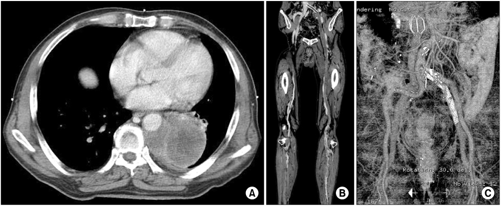
Fig. 1 Chest CT shows a 7.8 cm heterogeneously-enhancing mass in the medial aspect of the left lower lobe (A). Lower extremity CT angiogram reveals deep vein thrombosis involving the left lower extremity from the infrapopliteal veins to the common iliac vein (CIV), with a collapsed junction between the CIV and inferior vena cava (IVC) (B, C). IVC filter and vascular stent insertion on the left CIV was performed successfully.
- Full Text Links
-
- Actions
-
Cited
- CITED
-
- Close
- Share
-
- Similar articles
-
- May–Thurner Syndrome after Total Knee Arthroplasty
- May-Thurner Syndrome Appearing as Recurrent Swelling and Cellulitis in the Left Leg and Foot
- Iatrogenic Iliac Vein Injury Following Extracorporeal Membrane Oxygenation Cannulation in a Patient with May-Thurner Syndrome: A Case Report and Literature Review
- Ilio-Iliac Arteriovenous Fistula with May-Thurner Syndrome: A Case Report
- CT Findings of May–Thurner Syndrome in Diffuse Idiopathic Skeletal Hyperostosis: A Case Report
