Cervical design effect of dental implant on stress distribution in crestal cortical bone studied by finite element analysis
- Affiliations
-
- 1Department of Prosthodontics, School of Dentistry, Kyungpook National University, Korea. kblee@knu.ac.kr
- 2Department of Orthodontics, School of Dentistry, Kyungpook National University, Korea.
- KMID: 2195587
- DOI: http://doi.org/10.4047/jkap.2009.47.4.385
Abstract
- STATEMENT OF PROBLEM: High stress concentration on the crestal cortical bone has been regraded as a major etiologic factor jeopardizing long term stability of endosseous implants.
PURPOSE: To investigate if the design characteristics of crestal module, i.e. internal type, external type, and submerged type, affect stress distribution on the crestal cortical bone.
MATERIAL AND METHODS: A cylindrical shaped implant, 4.3 mm in diameter and 10 mm in length, with 3 different crestal modules, i.e. internal type, external type, and submerged type, were analysed. An axisymmetric scheme was used for finite elment formulation. A vertical load of 50 N and an oblique load of 50 N acting at 45degrees with the implant's long axis was applied. The peak crestal bone stress acting at the intersection of implant and crestal bone was compared.
RESULTS
Under vertical load, the crestal bone stress was high in the order of internal, external, and submerged types. Under the oblique loading condition, it was in the order of internal, submerged, and external types. CONCLUSION: Crestal module design was found to affect the level of the crestal bone stresses although the actual amount was not significant.
Figure
-
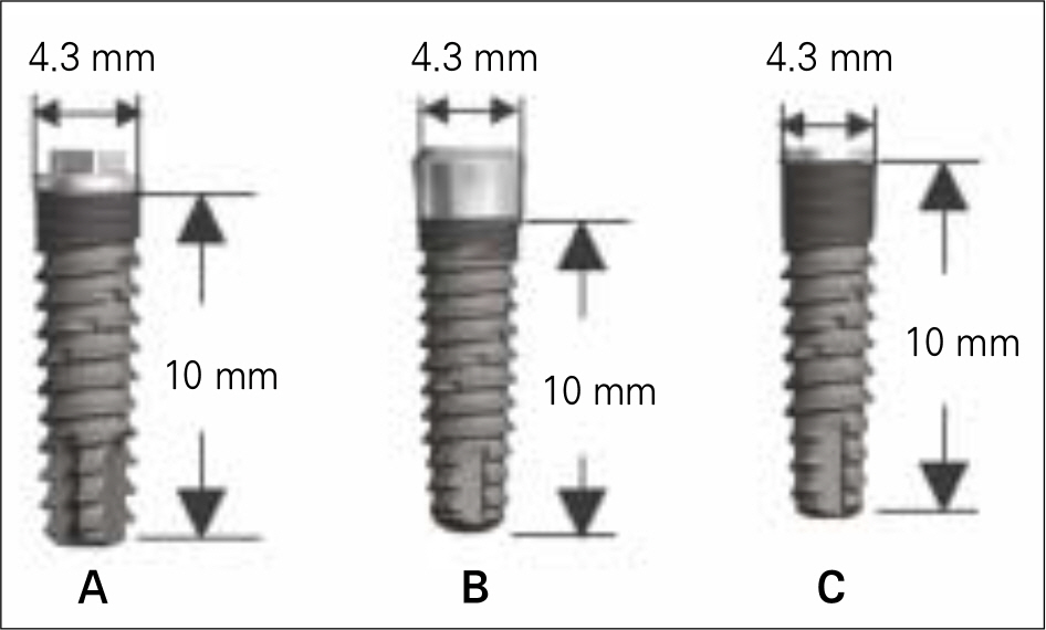
Fig. 1. Design configuration of 3 implants; A: external type, B: internal type, C: submerged type.

Fig. 2. Coordinate system and typical axisymmetric finite element model of the implant/bone complex subject to a load of 50 N acting at 45 degree with the implant axis. Soft tissues are not included in the F. E. model.
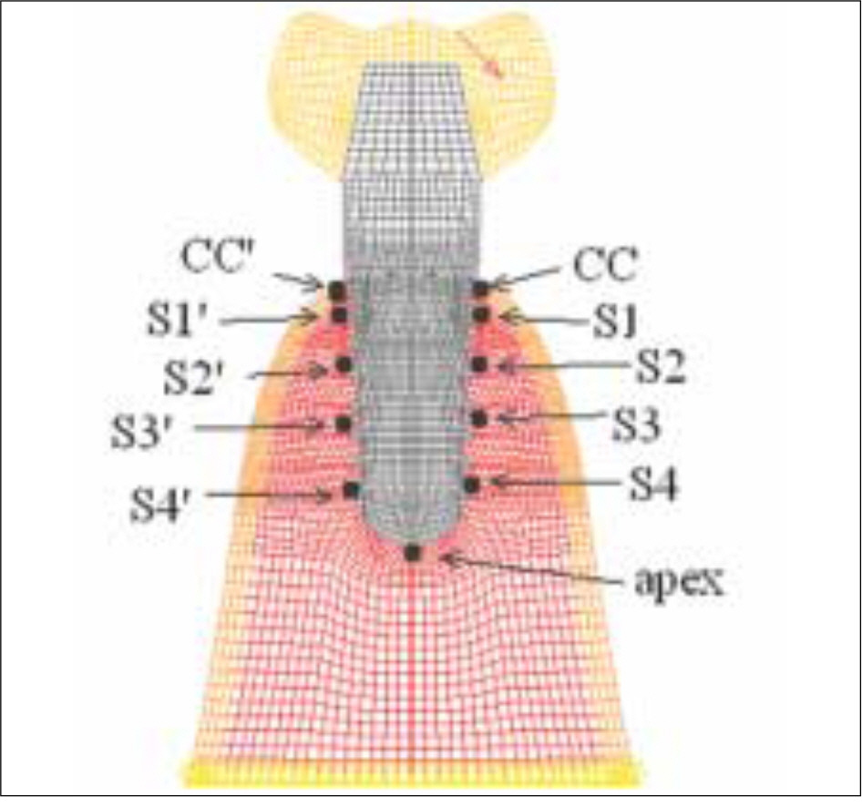
Fig. 3. Stress monitoring points in the bone: two points on the mid plane of cervical cortical plate, nine points in the cancellous bone.

Fig. 4. Maximum compressive stresses in the case of external type implant under: A: vertical load of 50 N, B: oblique load of 50 N.

Fig. 5. Magnification of the cervical area where the highesr stress concentration is noted: in the cases of A: vertical load, B: oblique load.

Fig. 6. Magnification of the cervical of the internal type implant subject to : A: vertical load of 50 N, B: oblique load of 50 N.

Fig. 7. Magnification of the cervical of the submerged type implant subject to : A: vertical load of 50 N, B: oblique load of 50 N.

Fig. 8. Stresses recorded at the 11 stress monitoring points under vertical load condition (50 N).

Fig. 9. Stresses recorded at the 11 stress monitoring points under oblique load condition (50 N).
Cited by 1 articles
-
Finite element analysis on the connection types of abutment and fixture
Byeong-Hyeon Jung, Gyeong-Je Lee, Dong-Wan Kang
J Korean Acad Prosthodont. 2012;50(2):119-127. doi: 10.4047/jkap.2012.50.2.119.
Reference
-
1.Becker W., Becker BE., Newman MG., Nyman S. Clinical and microbiologic findings that may contribute to dental implant failure. Int J Oral Maxillofac Implants. 1990. 5:31–8.2.Lindhe J., Berglundh T., Ericsson I., Liljenberg B., Marinello C. Experimental breakdown of peri-implant and periodontal tissues. A study in the beagle dog. Clin Oral Implants Res. 1992. 3:9–16.
Article3.Schou S., Holmstrup P., Reibel J., Juhl M., Hj � rting-Hansen E., Kornman KS. Ligature-induced marginal inflammation around osseointegrated implants and ankylosed teeth: stere-ologic and histologic observations in cynomolgus monkeys (Macaca fascicularis). J Periodontol. 1993. 64:529–37.
Article4.Vaillancourt H., Pilliar RM., McCammond D. Factors affecting crestal bone loss with dental implants partially covered with a porous coating: a finite element analysis. Int J Oral Maxillofac Implants. 1996. 11:351–9.5.Jung ES., Jo KH., Lee CH. A finite element stress analysis of the bone around implant following cervical bone resorption, J Korean Acad Implant Dent. 2003. 22:38–47.6.Kitamura E., Stegaroiu R., Nomura S., Miyakawa O. Influence of marginal bone resorption on stress around an implant - a three - dimensional finite element analysis. J Oral Rehabil. 2005. 32:279–86.7.Adell R., Lekholm U., Rockler B., Bra � nemark PI., Lindhe J., Eriksson B., Sbordone L. Marginal tissue reactions at osseointegrated titanium fixtures (I). A 3-year longitudinal prospective study. Int J Oral Maxillofac Surg. 1986. 15:39–52.8.Jemt T., Lekholm U., Adell R. Osseointegrated implants in the treatment of partially edentulous patients: a preliminary study on 876 consecutively placed fixtures. Int J Oral Maxillofac Implants. 1989. 4:211–7.9.Quirynen M., Naert I., van Steenberghe D. Fixture design and overload influence marginal bone loss and fixture success in the Bra � nemark system. Clin Oral Implants Res. 1992. 3:104–11.10.Hoshaw SJ., Brunski JB., Cochran GVB. Mechanical loading of Bra � nemark implants affects interfacial bone modeling and remodeling. Int J Oral & Maxillofac Implants. 1994. 9:345–60.11.Isidor F. Loss of osseointegration caused by occlusal load of oral implants. A clinical and radiographic study in monkeys. Clin Oral Implants Res. 1996. 7:143–52.
Article12.Isidor F. Histological evaluation of peri-implant bone at implants subjected to occlusal overload or plaque accumulation. Clin Oral Implants Res. 1997. 8:1–9.
Article13.Holmgren EP., Seckinger RJ., Kilgren LM., Mante F. Evaluating parameters of osseointegrated dental implants using finite element analysis—a two-dimensional comparative study examining the effects of implant diameter, implant shape, and load direction. J Oral Implantol. 1998. 24:80–8.
Article14.Van Oosterwyck H., Duyck J., Vander Sloten J., Van der Perre G., De Cooman M., Lievens S., Puers R., Naert I. The influence of bone mechanical properties and implant fixation upon bone loading around oral implants. Clin Oral Implants Res. 1998. 9:407–18.
Article15.O'Mahony AM., Williams JL., Spencer P. Anisotropic elasticity of cortical and cancellous bone in the posterior mandible increases peri-implant stress and strain under oblique loading. Clin Oral Implants Res. 2001. 12:648–57.16.Kum YJ. Finite Element Analysis of the Influences of the Implant Diameter on the Cortical Bone Stresses. MD thesis, Department of Dentistry, Kyungpook Nat Univ. 2004.17.Lim JY., Lee CH., Jo KH. A Finite Element Stress Analysis of the Bone aroung Implant following the Shape of Root Form Implant. J Korean Acad Implant Dent. 2003. 22:25–37.18.Lee JW., Lee CH., Jo KH. Finite element analysis of the stress distribution with load transfer characteristics of the implant/bone interface. J Korean Acad Implant Dent. 2003. 22:49–56.19.Lee SH. Stress analysis with nonlinear modelling of the load transfer characteristics across the osseointegrated interfaces of dental implant. MD thesis, Department of Dentistry, Kyungpook Nat Univ. 2002.20.Cha SB. Finite element approach to investigate the influence of the design configuration of the ITI solid implant on the bone stresses during the osseointegration process. MD thesis, Department of Dentistry, Kyungpook Nat Univ. 2005.21.Chang JM. Finite Element Approach to Investigate the Influence of the Jaw Bone Dimension on the Stresses Around the Root Analogue Dental Implant. MD thesis, Department of Dentistry, Kyungpook Nat Univ. 2004.22.Park DY. Three dimensional Stress Analysis Around Osseointegrated Bra � nemark Implant System Using An Axisymmetric Modelling Approach. MD Thesis, Kyungpook Nat Univ. 2001.23.Matsushita Y., Kitoh M., Mizuta K., Ikeda H., Suetsugu T. Two-dimensional FEM analysis of hydroxyapatite implants: diameter effects on stress distribution. J Oral Implantol. 1990. 16:6–11.24.Meijer HJ., Kuiper JH., Starmans FJ., Bosman F. Stress distribution around dental implants: influence of superstructure, length of implants, and height of mandible. J Prosthet Dent. 1992. 68:96–102.
Article25.Duyck J., Naert IE., Van Oosterwyck H., Van der Sloten J., De Cooman M., Lievens S., Puers B. Biomechanics of oral implants: a review of the literature. Technol Health Care. 1997. 5:253–73.
Article26.Palmer RM., Smith BJ., Palmer PJ., Floyd PD. A prospective study of Astra single tooth implants. Clin Oral Implants Res. 1997. 8:173–9.
Article27.Nordin T., Jo ¨nsson G., Nelvig P., Rasmusson L. The use of a conical fixture design for fixed partial prostheses. A preliminary report. Clin Oral Implants Res. 1998. 9:343–7.
Article28.Norton MR. Marginal bone levels at single tooth implants with a conical fixture design. The influence of surface macro - and microstructure. Clin Oral Implants Res. 1998. 9:91–9.29.Chun HJ., Cheong SY., Han JH., Heo SJ., Chung JP., Rhyu IC., Choi YC., Baik HK., Ku Y., Kim MH. Evaluation of design parameters of osseointegrated dental implants using finite element analysis. J Oral Rehabil. 2002. 29:565–74.
Article30.Tada S., Stegaroiu R., Kitamura E., Miyakawa O., Kusakari H. Influence of implant design and bone quality on stress/strain distribution in bone around implants: a 3-dimensional finite element analysis. Int J Oral Maxillofac Implants. 2003. 18:357–68.31.Lekholm U., Zarb GA. Patient selection and preparation. Bra � nemark PI, Zarb GA, Albrektsson T, editors. Tissue-integrated prostheses: osseointegration in clinical dentistry. Chicago: Quintessence;1985. p. 199–209.32.Asaoka K., Kuwayama N., Okuno O., Miura I. Mechanical properties and biomechanical compatibility of porous titanium for dental implants. J Biomed Mater Res. 1985. 19:699–713.
Article33.Papavasiliou G., Kamposiora P., Bayne SC., Felton DA. Three-dimensional finite element analysis of stress-distribution around single tooth implants as a function of bony support, prosthesis type, and loading during function. J Prosthet Dent. 1996. 76:633–40.
Article34.Richter EJ. In vivo vertical forces on implants. Int J Oral Maxillofac Implants. 1995. 10:99–108.35.Yu W., Jang YJ., Kyung HM. Combined influence of implant diameter and alveolar ridge width on crestal bone stress: a quantitative approach. Int J Oral Maxillofac Implants. 2009. 24:88–95.36.Frost HM. Bone's mechanostat: a 2003 update. Anat Rec. 2003. 275A:1081–101.
Article37.Meyer U., Joos U., Mythili J., Stamm T., Hohoff A., Stratmann U., Wiesmann HP. Ultrastructural characterization of the implant/bone interface of immediately loaded dental implants. Biometerials. 2004. 25:1959–67.
Article38.Hansson S., Werke M. The implant thread as a retention element in cortical bone: the effect of thread size and thread profile: a finite element study. J Biomech. 2003. 36:1247–58.
Article39.Isidor F. Loss of osseointegration caused by occlusal load of oral implants. A clinical and radiographic study in monkeys. Clin Oral Impl Res. 1996. 7:143–52.
Article
- Full Text Links
-
- Actions
-
Cited
- CITED
-
- Close
- Share
-
- Similar articles
-
- The effects of bone density and crestal cortical bone thickness on micromotion and peri-implant bone strain distribution in an immediately loaded implant: a nonlinear finite element analysis
- Finite element analysis of peri-implant bone stress influenced by cervical module configuration of endosseous implant
- THREE-DIMENSIONAL FINITE ELEMENT ANALYSIS OF THE EFFECT OF CORTICAL ENGAGEMENT ON IMPLANT LOAD TRANSFER IN POSTERIOR MANDIBLE
- An evaluation of angles between the alveolar crest bone and the implant effect on the implant crestal area induced stresses using a finite element method
- The effect of implant system with reverse beveled platform design on marginal bone stress distribution
