Biomechanical three-dimensional finite element analysis of monolithic zirconia crown with different cement type
- Affiliations
-
- 1Department of Dentistry, Ajou University School of Medicine, Suwon, Republic of Korea. dragon_001@hanmail.net
- KMID: 2176623
- DOI: http://doi.org/10.4047/jap.2015.7.6.475
Abstract
- PURPOSE
The objective of this study was to evaluate the influence of various cement types on the stress distribution in monolithic zirconia crowns under maximum bite force using the finite element analysis.
MATERIALS AND METHODS
The models of the prepared #46 crown (deep chamfer margin) were scanned and solid models composed of the monolithic zirconia crown, cement layer, and prepared tooth were produced using the computer-aided design technology and were subsequently translated into 3-dimensional finite element models. Four models were prepared according to different cement types (zinc phosphate, polycarboxylate, glass ionomer, and resin). A load of 700 N was applied vertically on the crowns (8 loading points). Maximum principal stress was determined.
RESULTS
Zinc phosphate cement had a greater stress concentration in the cement layer, while polycarboxylate cement had a greater stress concentration on the distal surface of the monolithic zirconia crown and abutment tooth. Resin cement and glass ionomer cement showed similar patterns, but resin cement showed a lower stress distribution on the lingual and mesial surface of the cement layer.
CONCLUSION
The test results indicate that the use of different luting agents that have various elastic moduli has an impact on the stress distribution of the monolithic zirconia crowns, cement layers, and abutment tooth. Resin cement is recommended for the luting agent of the monolithic zirconia crowns.
MeSH Terms
Figure
-
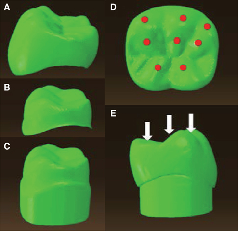
Fig. 1 Three components of the finite element analysis model. (A) Monolithic zirconia crown, (B) cement layer, (C) prepared tooth. Eight loading points and directions of load applying. (D), (E) three points on the outer inclines of each buccal cusp, three points on the inner inclines of each buccal cusp, and two points on the inner inclines of each lingual cusp.
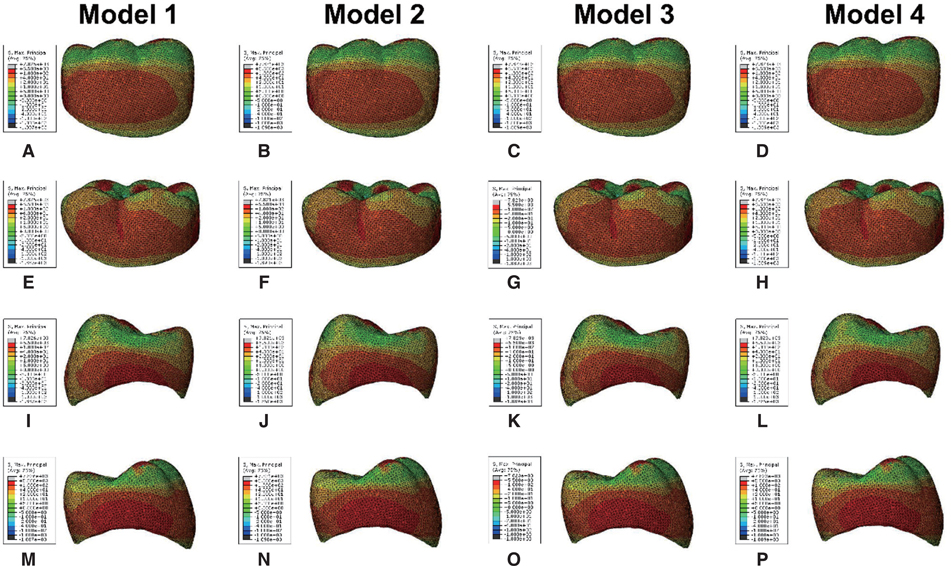
Fig. 2 Maximum principal stress distribution of the monolithic zirconia crown. (A) - (D) Buccal, (E) - (H) lingual, (I) - (L) mesial, (M) - (P) distal view.
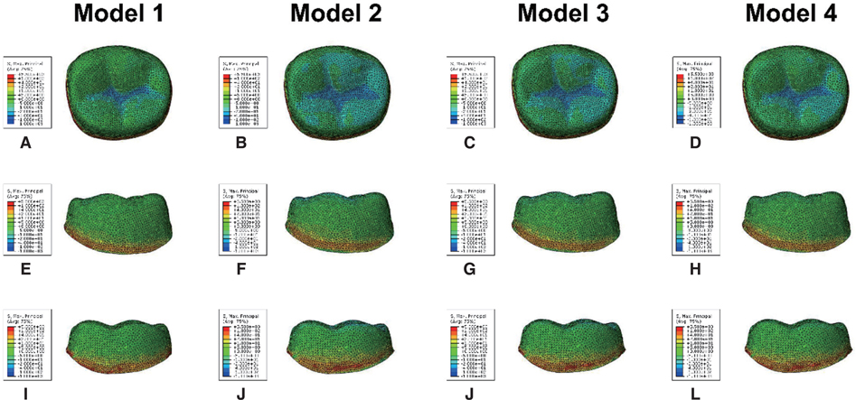
Fig. 3 Maximum principal stress distribution of the cement layer. (A) - (D) Occlusal, (E) - (H) buccal, (I) - (L) lingual view.
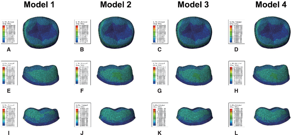
Fig. 4 Minimum principal stress distribution of the cement layer. (A) - (D) Occlusal, (E) - (H) buccal, (I) - (L) lingual view.
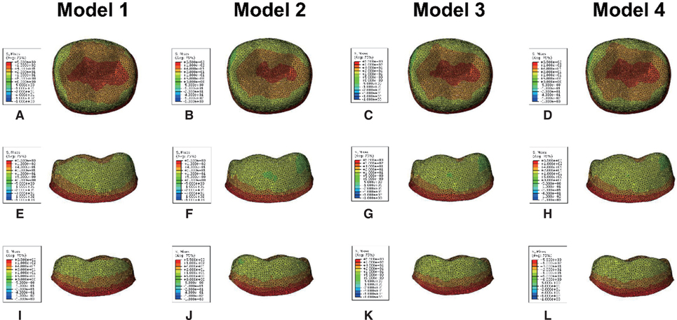
Fig. 5 von Mises stress distribution of the cement layer. (A) - (D) Occlusal, (E) - (H) buccal, (I) - (L) lingual view.
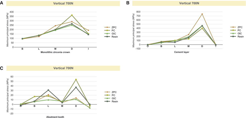
Fig. 6 The peak maximum principal stress value in each model. (A) Monolithic zirconia crown, (B) cement layer, (C) abutment tooth.

Fig. 7 The peak minimum principal stress value in each model. (A) Monolithic zirconia crown, (B) cement layer, (C) abutment tooth.

Fig. 8 The peak von Mises stress value in each model. (A) Monolithic zirconia crown, (B) cement layer, (C) abutment tooth.
Cited by 1 articles
-
In-vitro performance and fracture strength of thin monolithic zirconia crowns
Paul Weigl, Anna Sander, Yanyun Wu, Roland Felber, Hans-Christoph Lauer, Martin Rosentritt
J Adv Prosthodont. 2018;10(2):79-84. doi: 10.4047/jap.2018.10.2.79.
Reference
-
1. De Jager N, Pallav P, Feilzer AJ. The influence of design parameters on the FEA-determined stress distribution in CADCAM produced all-ceramic dental crowns. Dent Mater. 2005; 21:242–251.2. Piconi C, Maccauro G. Zirconia as a ceramic biomaterial. Biomaterials. 1999; 20:1–25.3. Shimizu K, Oka M, Kumar P, Kotoura Y, Yamamuro T, Makinouchi K, Nakamura T. Time-dependent changes in the mechanical properties of zirconia ceramic. J Biomed Mater Res. 1993; 27:729–734.4. Luthardt RG, Holzhüter M, Sandkuhl O, Herold V, Schnapp JD, Kuhlisch E, Walter M. Reliability and properties of ground Y-TZP-zirconia ceramics. J Dent Res. 2002; 81:487–491.5. Blatz MB, Sadan A, Martin J, Lang B. In vitro evaluation of shear bond strengths of resin to densely-sintered high-purity zirconium-oxide ceramic after long-term storage and thermal cycling. J Prosthet Dent. 2004; 91:356–362.6. Guess PC, Zavanelli RA, Silva NR, Bonfante EA, Coelho PG, Thompson VP. Monolithic CAD/CAM lithium disilicate versus veneered Y-TZP crowns: comparison of failure modes and reliability after fatigue. Int J Prosthodont. 2010; 23:434–442.7. McLaren EA, Terry DA. CAD/CAM systems, materials, and clinical guidelines for all-ceramic crowns and fixed partial dentures. Compend Contin Educ Dent. 2002; 23:637–641. 644646 passimquiz 654.8. Luthardt RG, Sandkuhl O, Herold V, Walter MH. Accuracy of mechanical digitizing with a CAD/CAM system for fixed restorations. Int J Prosthodont. 2001; 14:146–151.9. Preis V, Behr M, Hahnel S, Handel G, Rosentritt M. In vitro failure and fracture resistance of veneered and full-contour zirconia restorations. J Dent. 2012; 40:921–928.10. Ha SR, Kim SH, Han JS, Yoo SH, Jeong SC, Lee JB, Yeo IS. The influence of various core designs on stress distribution in the veneered zirconia crown: a finite element analysis study. J Adv Prosthodont. 2013; 5:187–197.11. Li X, Cao Z, Qiu X, Tang Z, Gong L, Wang D. Does matching relation exist between the length and the tilting angle of terminal implants in the all-on-four protocol? stress distributions by 3D finite element analysis. J Adv Prosthodont. 2015; 7:240–248.12. Spazzin AO, Galafassi D, de Meira-Júnior AD, Braz R, Garbin CA. Influence of post and resin cement on stress distribution of maxillary central incisors restored with direct resin composite. Oper Dent. 2009; 34:223–229.13. Soares CJ, Raposo LH, Soares PV, Santos-Filho PC, Menezes MS, Soares PB, Magalhães D. Effect of different cements onthe biomechanical behavior of teeth restored with cast dowel- and-cores-in vitro and FEA analysis. J Prosthodont. 2010; 19:130–137.14. Pilo R, Cardash HS. In vivo retrospective study of cement thickness under crowns. J Prosthet Dent. 1998; 79:621–625.15. Reich S, Wichmann M, Nkenke E, Proeschel P. Clinical fit of all-ceramic three-unit fixed partial dentures, generated with three different CAD/CAM systems. Eur J Oral Sci. 2005; 113:174–179.16. Proos KA, Swain MV, Ironside J, Steven GP. Influence of cement on a restored crown of a first premolar using finite element analysis. Int J Prosthodont. 2003; 16:82–90.17. Ferrario VF, Sforza C, Zanotti G, Tartaglia GM. Maximal bite forces in healthy young adults as predicted by surface electromyography. J Dent. 2004; 32:451–457.18. Nishigawa K, Bando E, Nakano M. Quantitative study of bite force during sleep associated bruxism. J Oral Rehabil. 2001; 28:485–491.19. Manso AP, Silva NR, Bonfante EA, Pegoraro TA, Dias RA, Carvalho RM. Cements and adhesives for all-ceramic restorations. Dent Clin North Am. 2011; 55:311–332, ix.20. Palacios RP, Johnson GH, Phillips KM, Raigrodski AJ. Retention of zirconium oxide ceramic crowns with three types of cement. J Prosthet Dent. 2006; 96:104–114.21. Blatz MB, Sadan A, Kern M. Resin-ceramic bonding: a review of the literature. J Prosthet Dent. 2003; 89:268–274.22. Ernst CP, Cohnen U, Stender E, Willershausen B. In vitro retentive strength of zirconium oxide ceramic crowns using different luting agents. J Prosthet Dent. 2005; 93:551–558.23. Pospiech P. All-ceramic crowns: bonding or cementing? Clin Oral Investig. 2002; 6:189–197.24. Thompson JY, Stoner BR, Piascik JR, Smith R. Adhesion/cementation to zirconia and other non-silicate ceramics: where are we now? Dent Mater. 2011; 27:71–82.25. Kamposiora P, Papavasilious G, Bayne SC, Felton DA. Finite element analysis estimates of cement microfracture under complete veneer crowns. J Prosthet Dent. 1994; 71:435–441.26. Shahrbaf S, vanNoort R, Mirzakouchaki B, Ghassemieh E, Martin N. Effect of the crown design and interface lute parameters on the stress-state of a machined crown-tooth system: a finite element analysis. Dent Mater. 2013; 29:e123–e131.27. Rekow ED, Harsono M, Janal M, Thompson VP, Zhang G. Factorial analysis of variables influencing stress in all-ceramic crowns. Dent Mater. 2006; 22:125–132.28. May LG, Kelly JR, Bottino MA, Hill T. Effects of cement thickness and bonding on the failure loads of CAD/CAM ceramic crowns: multi-physics FEA modeling and monotonic testing. Dent Mater. 2012; 28:e99–109.29. Ayad MF, Rosenstiel SF, Salama M. Influence of tooth surface roughness and type of cement on retention of complete cast crowns. J Prosthet Dent. 1997; 77:116–121.30. Pameijer CH. A review of luting agents. Int J Dent. 2012; 2012:752861.
- Full Text Links
-
- Actions
-
Cited
- CITED
-
- Close
- Share
-
- Similar articles
-
- An evaluation of the stress effect of different occlusion concepts on hybrid abutment and implant supported monolithic zirconia fixed prosthesis: A finite element analysis
- Fracture strength of zirconia monolithic crowns
- Finite element analysis of the influence of esthetic posts on incisors
- Evaluation of reliability of zirconia materials to be used in implant-retained restoration on the atrophic bone of the posterior maxilla: A finite element study
- Three-dimensional finite element analysis of implant-supported crown in fibula bone model
