J Adv Prosthodont.
2013 Aug;5(3):326-332. 10.4047/jap.2013.5.3.326.
Three-dimensional finite element analysis of implant-supported crown in fibula bone model
- Affiliations
-
- 1Department of Oral Anatomy and Dental Research Institute, School of Dentistry, Seoul National University, Seoul, Republic of Korea.
- 2Department of Prosthodontics and Dental Research Institute, School of Dentistry, Seoul National University, Seoul, Republic of Korea. proskwon@snu.ac.kr
- KMID: 2118173
- DOI: http://doi.org/10.4047/jap.2013.5.3.326
Abstract
- PURPOSE
The purpose of this study was to compare stress distributions of implant-supported crown placed in fibula bone model with those in intact mandible model using three-dimensional finite element analysis.
MATERIALS AND METHODS
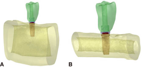
Two three-dimensional finite element models were created to analyze biomechanical behaviors of implant-supported crowns placed in intact mandible and fibula model. The finite element models were generated from patient's computed tomography data. The model for grafted fibula was composed of fibula block, dental implant system, and implant-supported crown. In the mandible model, same components with identical geometries with the fibula model were used except that the mandible replaced the fibula. Vertical and oblique loadings were applied on the crowns. The highest von Mises stresses were investigated and stress distributions of the two models were analyzed.
RESULTS
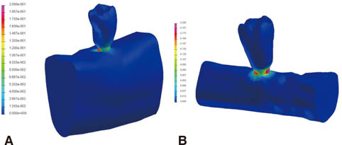
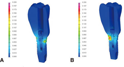
Overall stress distributions in the two models were similar. The highest von Mises stress values were higher in the mandible model than in the fibula model. In the individual prosthodontic components there was no prominent difference between models. The stress concentrations occurred in cortical bones in both models and the effect of bicortical anchorage could be found in the fibula model.
CONCLUSION
Using finite element analysis it was shown that the implant-supported crown placed in free fibula graft might function successfully in terms of biomechanical behavior.
MeSH Terms
Figure
Reference
-
1. Beumer J III, Marunick MT, Esposito SJ. Maxillofacial Rehabilitation: Surgical and Prosthodontic Management of Cancer-Related Acquired, and Congenital Defects of the Head and Neck. 1st ed. Chicago: Quintessence Publishing Co, Inc.;2011. p. 61–154.2. Olson ML, Shedd DP. Disability and rehabilitation in head and neck cancer patients after treatment. Head Neck Surg. 1978; 1:52–58.3. Komisar A. The functional result of mandibular reconstruction. Laryngoscope. 1990; 100:364–374.4. Hidalgo DA. Fibula free flap: a new method of mandible reconstruction. Plast Reconstr Surg. 1989; 84:71–79.5. Ferrari S, Bianchi B, Savi A, Poli T, Multinu A, Balestreri A, Ferri A. Fibula free flap with endosseous implants for reconstructing a resected mandible in bisphosphonate osteonecrosis. J Oral Maxillofac Surg. 2008; 66:999–1003.6. Zlotolow IM, Huryn JM, Piro JD, Lenchewski E, Hidalgo DA. Osseointegrated implants and functional prosthetic rehabilitation in microvascular fibula free flap reconstructed mandibles. Am J Surg. 1992; 164:677–681.7. Hayter JP, Cawood JI. Oral rehabilitation with endosteal implants and free flaps. Int J Oral Maxillofac Surg. 1996; 25:3–12.8. Lukash FN, Sachs SA, Fischman B, Attie JN. Osseointegrated denture in a vascularized bone transfer: functional jaw reconstruction. Ann Plast Surg. 1987; 19:538–544.9. Hidalgo DA. Aesthetic improvements in free-flap mandible reconstruction. Plast Reconstr Surg. 1991; 88:574–585.10. Gürlek A, Miller MJ, Jacob RF, Lively JA, Schusterman MA. Functional results of dental restoration with osseointegrated implants after mandible reconstruction. Plast Reconstr Surg. 1998; 101:650–655.11. Urken ML, Buchbinder D, Weinberg H, Vickery C, Sheiner A, Parker R, Schaefer J, Som P, Shapiro A, Lawson W, et al. Functional evaluation following microvascular oromandibular reconstruction of the oral cancer patient: a comparative study of reconstructed and nonreconstructed patients. Laryngoscope. 1991; 101:935–950.12. Frodel JL Jr, Funk GF, Capper DT, Fridrich KL, Blumer JR, Haller JR, Hoffman HT. Osseointegrated implants: a comparative study of bone thickness in four vascularized bone flaps. Plast Reconstr Surg. 1993; 92:449–455.13. Horiuchi K, Hattori A, Inada I, Kamibayashi T, Sugimura M, Yajima H, Tamai S. Mandibular reconstruction using the double barrel fibular graft. Microsurgery. 1995; 16:450–454.14. Chang YM, Santamaria E, Wei FC, Chen HC, Chan CP, Shen YF, Hou SP. Primary insertion of osseointegrated dental implants into fibula osteoseptocutaneous free flap for mandible reconstruction. Plast Reconstr Surg. 1998; 102:680–688.15. Marunick MT, Roumanas ED. Functional criteria for mandibular implant placement post resection and reconstruction for cancer. J Prosthet Dent. 1999; 82:107–113.16. Esposito M, Hirsch JM, Lekholm U, Thomsen P. Biological factors contributing to failures of osseointegrated oral implants. (II). Etiopathogenesis. Eur J Oral Sci. 1998; 106:721–764.17. van Eijden TM. Biomechanics of the mandible. Crit Rev Oral Biol Med. 2000; 11:123–136.18. Geng JP, Tan KB, Liu GR. Application of finite element analysis in implant dentistry: a review of the literature. J Prosthet Dent. 2001; 85:585–598.19. Meijer HJ, Kuiper JH, Starmans FJ, Bosman F. Stress distribution around dental implants: influence of superstructure, length of implants, and height of mandible. J Prosthet Dent. 1992; 68:96–102.20. Baggi L, Pastore S, Di Girolamo M, Vairo G. Implant-bone load transfer mechanisms in complete-arch prostheses supported by four implants: a three-dimensional finite element approach. J Prosthet Dent. 2013; 109:9–21.21. de Paula GA, da Mota AS, Moreira AN, de Magahlães CS, Cornacchia TP, Cimini CA Jr. The effect of prosthesis length and implant diameter on the stress distribution in tooth-implant-supported prostheses: a finite element analysis. Int J Oral Maxillofac Implants. 2012; 27:e19–e28.22. Wang RF, Kang B, Lang LA, Razzoog ME. The dynamic natures of implant loading. J Prosthet Dent. 2009; 101:359–371.23. Wu JC, Chen CS, Yip SW, Hsu ML. Stress distribution and micromotion analyses of immediately loaded implants of varying lengths in the mandible and fibular bone grafts: a three-dimensional finite element analysis. Int J Oral Maxillofac Implants. 2012; 27:e77–e84.24. Rho JY, Ashman RB, Turner CH. Young's modulus of trabecular and cortical bone material: ultrasonic and microtensile measurements. J Biomech. 1993; 26:111–119.25. Haraldson T, Carlsson GE. Bite force and oral function in patients with osseointegrated oral implants. Scand J Dent Res. 1977; 85:200–208.26. Liao SH, Tong RF, Dong JX. Anisotropic finite element modeling for patient-specific mandible. Comput Methods Programs Biomed. 2007; 88:197–209.27. Peled M, El-Naaj IA, Lipin Y, Ardekian L. The use of free fibular flap for functional mandibular reconstruction. J Oral Maxillofac Surg. 2005; 63:220–224.28. Hidalgo DA, Rekow A. A review of 60 consecutive fibula free flap mandible reconstructions. Plast Reconstr Surg. 1995; 96:585–596.29. Sieg P, Zieron JO, Bierwolf S, Hakim SG. Defect-related variations in mandibular reconstruction using fibula grafts. A review of 96 cases. Br J Oral Maxillofac Surg. 2002; 40:322–329.30. Smolka K, Kraehenbuehl M, Eggensperger N, Hallermann W, Thoren H, Iizuka T, Smolka W. Fibula free flap reconstruction of the mandible in cancer patients: evaluation of a combined surgical and prosthodontic treatment concept. Oral Oncol. 2008; 44:571–581.31. Kido H, Schulz EE, Kumar A, Lozada J, Saha S. Implant diameter and bone density: effect on initial stability and pull-out resistance. J Oral Implantol. 1997; 23:163–169.32. Myoung H, Kim YY, Heo MS, Lee SS, Choi SC, Kim MJ. Comparative radiologic study of bone density and cortical thickness of donor bone used in mandibular reconstruction. Oral Surg Oral Med Oral Pathol Oral Radiol Endod. 2001; 92:23–29.33. van Staden RC, Guan H, Loo YC. Application of the finite element method in dental implant research. Comput Methods Biomech Biomed Engin. 2006; 9:257–270.34. Chou HY, Müftü S, Bozkaya D. Combined effects of implant insertion depth and alveolar bone quality on periimplant bone strain induced by a wide-diameter, short implant and a narrow-diameter, long implant. J Prosthet Dent. 2010; 104:293–300.35. Holmes DC, Loftus JT. Influence of bone quality on stress distribution for endosseous implants. J Oral Implantol. 1997; 23:104–111.36. Brunski JB. Biomechanical factors affecting the bone-dental implant interface. Clin Mater. 1992; 10:153–201.37. Ichikawa T, Kanitani H, Wigianto R, Kawamoto N, Matsumoto N. Influence of bone quality on the stress distribution. An in vitro experiment. Clin Oral Implants Res. 1997; 8:18–22.38. Clelland NL, Lee JK, Bimbenet OC, Gilat A. Use of an axisymmetric finite element method to compare maxillary bone variables for a loaded implant. J Prosthodont. 1993; 2:183–189.39. Truhlar RS, Orenstein IH, Morris HF, Ochi S. Distribution of bone quality in patients receiving endosseous dental implants. J Oral Maxillofac Surg. 1997; 55:38–45.40. Huang HL, Fuh LJ, Ko CC, Hsu JT, Chen CC. Biomechanical effects of a maxillary implant in the augmented sinus: a three-dimensional finite element analysis. Int J Oral Maxillofac Implants. 2009; 24:455–462.41. Demenko V, Linetskiy I, Nesvit K, Shevchenko A. Ultimate masticatory force as a criterion in implant selection. J Dent Res. 2011; 90:1211–1215.42. van Oosterwyck H, Duyck J, van der Sloten J, van der Perre G, De Cooman M, Lievens S, Puers R, Naert I. The influence of bone mechanical properties and implant fixation upon bone loading around oral implants. Clin Oral Implants Res. 1998; 9:407–418.
- Full Text Links
- Actions
-
Cited
- CITED
-
- Close
- Share
- Similar articles
-
- Three-dimensional finite element analysis of the splinted implant prosthesis in a reconstructed mandible
- Three-dimensional finite element analysis of platform switched implant
- Effects of crown retrieval on implants and the surrounding bone: a finite element analysis
- Three-dimensional finite element analysis for influence of marginal bone resorption on stress distribution in internal conical joint type implant fixture
- The three dimensional finite element analysis of the stress distribution according to the thread designs and the marginal bone loss of the implants

