Investig Magn Reson Imaging.
2015 Sep;19(3):186-190. 10.13104/imri.2015.19.3.186.
Acute Disseminated Encephalomyelitis Presenting as Rhombencephalitis: An Atypical Case Presentation
- Affiliations
-
- 1Section of Neuroradiology, Department of Radiology, Soonchunhyang University Bucheon Hospital, Gyeonggi-do, Korea. aleerad@schmc.ac.kr
- KMID: 2175614
- DOI: http://doi.org/10.13104/imri.2015.19.3.186
Abstract
- Acute disseminated encephalomyelitis (ADEM) is a demyelinating and inflammatory condition of the central nervous system, occurring predominantly in white matter. ADEM involving the rhombencephalon without affecting the white matter is very rare. Here, we present an unusual case of ADEM involving only the rhombencephalon in a 4-year-old Asian girl. The patient complained of pain in the right lower extremities, general weakness, ataxia, and dysarthria. The initial brain CT showed subtle ill-defined low-density lesions in the pons and medulla. On brain MRI, T2 high signal intensity (T2-HSI) lesions with mild swelling were present in the pons, both middle cerebellar peduncles, and the anterior medulla. The initial diagnosis was viral encephalitis involving the rhombencephalon. Curiously, a cerebrospinal fluid (CSF) study revealed no cellularity, and negative viral marker findings. Three weeks later, follow up brain MRI showed that the extent of the T2-HSI lesions in the brain stem had decreased. After reinvestigation, it was found that she had a prior history of upper respiratory infection. In this case, we report the very rare case of a patient showing isolated involvement of the rhombencephalon in ADEM, mimicking viral rhombencephalitis on CT and MR imaging. ADEM can involve unusual sites such as the rhombencephalon in isolation, without involvement of the white matter or deep gray matter and, therefore, should be considered even when it appears in unusual anatomical areas. Thorough history taking is important for making a correct diagnosis.
Keyword
MeSH Terms
-
Asian Continental Ancestry Group
Ataxia
Biomarkers
Brain
Brain Stem
Central Nervous System
Cerebrospinal Fluid
Child, Preschool
Diagnosis
Dysarthria
Encephalitis, Viral
Encephalomyelitis
Encephalomyelitis, Acute Disseminated*
Female
Follow-Up Studies
Humans
Lower Extremity
Magnetic Resonance Imaging
Pons
Rhombencephalon
Figure
-
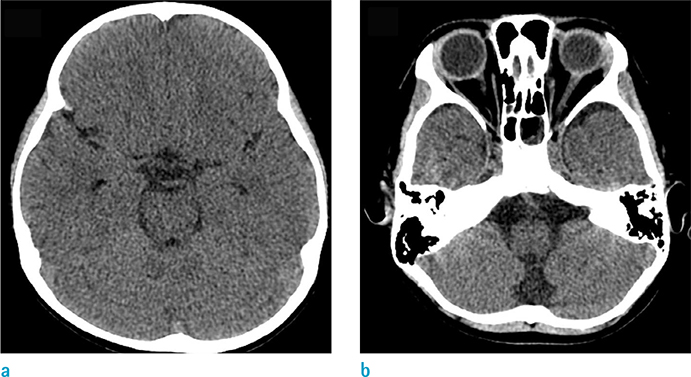
Fig. 1 Non-enhanced brain CT axial images (a, b) showing equivocal, ill-defined low-density lesions in the pons and medulla.
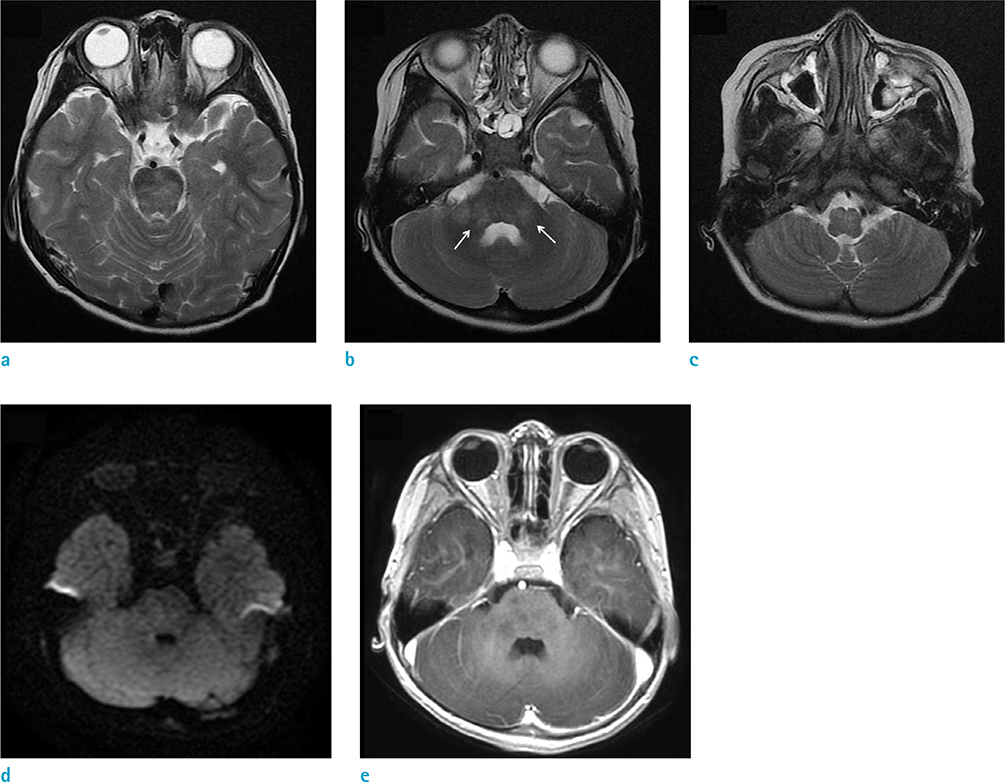
Fig. 2 Imaging findings of the lesion on brain MRI. (a-c) Axial T2WI showing ill-defined high signal intensity (HSI) with parenchymal swelling in the pons, both middle cerebellar peduncles (arrows), and anterior medulla. (d) Diffusion-weighted imaging showed no diffusion restriction at the sites of the T2-HSI lesions. (e) No definite lesion enhancement is seen on the MR image.
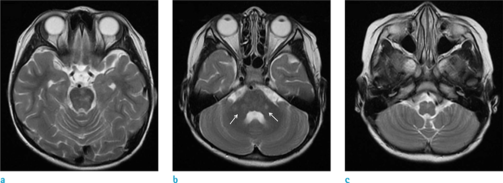
Fig. 3 Follow-up MRI scans at 40 days after the onset of clinical symptoms. (a-c) Axial T2-weighted images showing decreased number and extent of T2-HSI in the pons, both middle cerebellar peduncles (arrows), and the medulla.
Reference
-
1. Dale RC. Acute disseminated encephalomyelitis. Semin Pediatr Infect Dis. 2003; 14:90–95.2. Osborn AG. Osborn's brain: Imaging, pathology, and anatomy. 1st ed. Philadelphia, PA: Lippincott Williams & Wilkins;2012.3. Garg RK. Acute disseminated encephalomyelitis. Postgrad Med J. 2003; 79:11–17.4. Murthy SN, Faden HS, Cohen ME, Bakshi R. Acute disseminated encephalomyelitis in children. Pediatrics. 2002; 110:e21.5. Apak RA, Kose G, Anlar B, Turanli G, Topaloglu H, Ozdirim E. Acute disseminated encephalomyelitis in childhood: report of 10 cases. J Child Neurol. 1999; 14:198–201.6. Firat AK, Karakas HM, Yakinci C, Altinok T, Alkan A, Bicak U. An unusual case of acute disseminated encephalomyelitis confined to brainstem. Magn Reson Imaging. 2004; 22:1329–1332.7. Singh S, Alexander M, Korah IP. Acute disseminated encephalomyelitis: MR imaging features. AJR Am J Roentgenol. 1999; 173:1101–1107.8. Alper G, Sreedher G, Zuccoli G. Isolated brain stem lesion in children: is it acute disseminated encephalomyelitis or not? AJNR Am J Neuroradiol. 2013; 34:217–220.9. Tenembaum S, Chitnis T, Ness J, Hahn JS. International Pediatric MSSG. Acute disseminated encephalomyelitis. Neurology. 2007; 68:S23–S36.10. Mialin R, Koob M, de Seze J, Dietemann JL, Kremer S. Case 173: acute disseminated encephalomyelitis confined to the brainstem. Radiology. 2011; 260:911–914.
- Full Text Links
-
- Actions
-
Cited
- CITED
-
- Close
- Share
-
- Similar articles
-
- Acute disseminated encephalomyelitis
- A case of acute disseminated encephalomyelitis
- A Case of Multiple Sclerosis Presenting as Acute Disseminated Encephalomyelitis
- Acute Disseminated Encephalomyelitis Following Pneumococcal Vaccination
- Acute Disseminated Encephalomyelitis Associated with Influenza Vaccination
