Reduction of Inter-Rater and Intra-Rater Variability in Psoriasis Area and Severity Index Assessment by Photographic Training
- Affiliations
-
- 1Department of Dermatology, Seoul National University Bundang Hospital, Seoul National University College of Medicine, Seongnam, Korea. swyoun@snu.ac.kr
- KMID: 2171456
- DOI: http://doi.org/10.5021/ad.2015.27.5.557
Abstract
- BACKGROUND
Severity grading is important for the assessment of psoriasis treatment efficacy. This is most commonly achieved by using the psoriasis area and severity index (PASI), a subjective tool with inherent inter-rater and intra-rater variability. PASI-naive dermatologists require training to properly conduct a PASI assessment.
OBJECTIVE
In the present study, we aimed to investigate whether photographic training improves inter-rater and intra-rater variabilities. We also determined which PASI component has the greatest impact on variability.
METHODS
Twenty-one dermatologists received 1 hour of PASI training. They were tested before and after the training to evaluate intra-rater variability. The physicians were further tested after training by using a reference photograph.
RESULTS
The mean of each PASI component was underevaluated compared with scoring by a PASI expert. The concordance rate with the expert's grading was highest for thickness followed by erythema, scaling, and area. The scaling score showed the greatest improvement after training. After training, the distribution of deviation from the expert's grading, which signifies inter-rater variability, improved only for the PASI area component. The deviation of scaling grading improved upon retesting by using a reference photograph.
CONCLUSION
PASI assessment training improved variabilities to some degree but not for every PASI component. The development of an objective psoriasis severity assessment tool will help overcome the subjective variabilities in PASI assessment, which can never be completely eliminated via training.
Figure
-

Fig. 1 Comparisons of the grading results (A) between the pretest and follow-up retest for mean erythema, thickness, and scaling grades; (B) between the pretest and retest for mean area estimates (%); (C) between the pretest and retest with reference photographs for mean erythema, thickness, and scaling grades. *p<0.05.
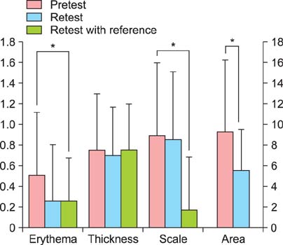
Fig. 2 Deviations from the expert's grading for the psoriasis area and severity index assessment components in three tests. The left side scale is the y-axis of erythema, thickness, and scaling (grading), and the right side scale is the y-axis of the area (%). *p<0.05.

Fig. 3 Coverage area (%) estimation module for psoriasis area and severity index body surface area calculation. When we slide the control bar under the rectangle, the area (%) covered by the circles in the main rectangle changes. The sum of the circle areas represents (A) 10% and (B) 33% coverage of the rectangle. To estimate the area covered by small plaques distributed across a body region, the approximate area (%) can be obtained by sliding the control bar to display a size similar to that of the patient's lesion in the main rectangle.
Cited by 5 articles
-
Recent medical therapy for psoriasis
Sang Woong Youn
J Korean Med Assoc. 2019;62(3):176-180. doi: 10.5124/jkma.2019.62.3.176.The MARCOPOLO Study of Ustekinumab Utilization and Efficacy in a Real-World Setting: Treatment of Patients with Plaque Psoriasis in Asia-Pacific Countries
Sang Woong Youn, Tsen-Fang Tsai, Colin Theng, Siew-Eng Choon, Benny E. Wiryadi, Antonio Pires, Weihao Tan, Min-Geol Lee,
Ann Dermatol. 2016;28(2):222-231. doi: 10.5021/ad.2016.28.2.222.Characteristics of Pruritus according to Morphological Phenotype of Psoriasis and Association with Neuropeptides and Interleukin-31
Sung-Min Park, Gun-Wook Kim, Hoon-Soo Kim, Hyun-Chang Ko, Moon-Bum Kim, Byung-Soo Kim
Ann Dermatol. 2020;32(1):1-7. doi: 10.5021/ad.2020.32.1.1.Both Educational Lectures and Reference Photographs Are Necessary to Improve the Accuracy and Reliability of Psoriasis Area and Severity Index (PASI) Assessment: Results from Korean Nation-Wide PASI Educational Workshop
Chong Won Choi, Bo Ri Kim, Jong Seo Park, Sang Woong Youn
Ann Dermatol. 2018;30(3):284-289. doi: 10.5021/ad.2018.30.3.284.Quantification of Efflorescences in Pustular Psoriasis Using Deep Learning
Ludovic Amruthalingam, Oliver Buerzle, Philippe Gottfrois, Alvaro Gonzalez Jimenez, Anastasia Roth, Thomas Koller, Marc Pouly, Alexander A. Navarini
Healthc Inform Res. 2022;28(3):222-230. doi: 10.4258/hir.2022.28.3.222.
Reference
-
1. Robinson A, Kardos M, Kimball AB. Physician Global Assessment (PGA) and Psoriasis Area and Severity Index (PASI): why do both? A systematic analysis of randomized controlled trials of biologic agents for moderate to severe plaque psoriasis. J Am Acad Dermatol. 2012; 66:369–375.
Article2. Kim BY, Choi JW, Kim BR, Youn SW. Histopathological findings are associated with the clinical types of psoriasis but not with the corresponding lesional psoriasis severity index. Ann Dermatol. 2015; 27:26–31.
Article3. Armstrong AW, Parsi K, Schupp CW, Mease PJ, Duffin KC. Standardizing training for psoriasis measures: effectiveness of an online training video on Psoriasis Area and Severity Index assessment by physician and patient raters. JAMA Dermatol. 2013; 149:577–582.4. van de Kerkhof PC. On the limitations of the psoriasis area and severity index (PASI). Br J Dermatol. 1992; 126:205.
Article5. Gelfand JM, Weinstein R, Porter SB, Neimann AL, Berlin JA, Margolis DJ. Prevalence and treatment of psoriasis in the United Kingdom: a population-based study. Arch Dermatol. 2005; 141:1537–1541.6. Faria JR, Aarão AR, Jimenez LM, Silva OH, Avelleira JC. Inter-rater concordance study of the PASI (Psoriasis Area and Severity Index). An Bras Dermatol. 2010; 85:625–629.7. Ramsay B, Lawrence CM. Measurement of involved surface area in patients with psoriasis. Br J Dermatol. 1991; 124:565–570.
Article8. Speight EL, Essex TJ, Farr PM. The study of plaques of psoriasis using a scanning laser-Doppler velocimeter. Br J Dermatol. 1993; 128:519–524.
Article9. Choi JW, Kwon SH, Youn JI, Youn SW. Objective measurements of erythema, elasticity and scale could overcome the inter- and intra-observer variations of subjective evaluations for psoriasis severity. Eur J Dermatol. 2013; 23:224–229.
Article10. Choi JW, Kim BR, Choi CW, Youn SW. One device, one equation: the simplest way to objectively evaluate psoriasis severity. J Dermatol. 2015; 42:154–158.
Article
- Full Text Links
-
- Actions
-
Cited
- CITED
-
- Close
- Share
-
- Similar articles
-
- Both Educational Lectures and Reference Photographs Are Necessary to Improve the Accuracy and Reliability of Psoriasis Area and Severity Index (PASI) Assessment: Results from Korean Nation-Wide PASI Educational Workshop
- Intra and Inter-Rater Measurement Reliability of Tibialis Anterior Muscle (TA) Thickness using the Ultrasonography Spring Gauge Technique
- The Intra- and Inter-rater Reliability and the Learning Curve for a Simple Neurological Score for Rats
- The Availability of Radiological Measurement of Femoral Anteversion Angle: Three-Dimensional Computed Tomography Reconstruction
- Extraction of the pull force from inertial sensors during the pull test for Parkinson’s disease: A reliability study
