Accelerated and Exacerbated Effects of High Dietary Fat on Neuronal Damage Induced by Transient Cerebral Ischemia in the Gerbil Septum
- Affiliations
-
- 1Department of Emergency Medicine, Kangwon National University School of Medicine, Chuncheon, Korea. cjhmd@kangwon.ac.kr
- 2Department of Integrative Traditional & Western Medicine, Yangzhou University Medical College, Yangzhou, China.
- 3Department of Physiology and Institute of Neurodegeneration and Neuroregeneration, Hallym University College of Medicine, Chuncheon, Korea.
- 4Department of Neurobiology, Kangwon National University School of Medicine, Chuncheon, Korea. mhwon@kangwon.ac.kr
- 5Department of Emergency Medicine, Yonsei University College of Medicine, Seoul, Korea.
- KMID: 2169470
- DOI: http://doi.org/10.3803/EnM.2014.29.3.328
Abstract
- BACKGROUND
Obesity induced by high-fat diet (HFD) is one of the most widespread metabolic disorders in current society. However, there has been little research regarding the effects of HFD-induced obesity in the septa of animal models of cerebral ischemia. Therefore, in the present study, we investigated septal effects of HFD on neuronal damage and gliosis induced by transient cerebral ischemia.
METHODS
Body weight, blood glucose levels and serum lipid profiles levels were measured both in the normal diet (ND) and HFD-group. We also investigated the effects of ND and HFD on neuronal damage and gliosis in the septum after transient cerebral ischemia using immunohistochemistry.
RESULTS
The levels of blood glucose, serum triglyceride, and total cholesterol were significantly increased in the HFD-fed gerbils compared with the ND-fed gerbils, although body weight was not significantly changed after HFD feeding. In the ND-fed gerbils, ischemia-induced neuronal damage was found in the septohippocampal nucleus (SHN) of the septum 7 days after ischemia. In the HFD-fed gerbils, ischemia-induced neuronal damage in the SHN was much more severe compared with that of the ND-fed gerbils 4 and 7 days after ischemia. In addition, we found that ischemia-induced glial activation including astrocytes and microglia was accelerated and exacerbated in the HFD-fed gerbils compared with that in the ND-fed gerbils.
CONCLUSION
These results indicate that HFD can lead to much more severe effects in ischemia-induced neuronal damage/death in the septum after ischemia-reperfusion, and that it may be associated with accelerated change in glial activation.
MeSH Terms
Figure
-
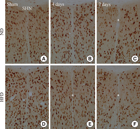
Fig. 1 (A-F) NeuN immunohistochemistry in the septohippocampal nucleus (SHN) of the septum in the normal diet (ND)- and high-fat diet (HFD)-groups at sham, 4 and 7 days after ischemia-reperfusion. A loss of NeuN+ neurons (asterisks) is detected 4 and 7 days after ischemia-reperfusion in the HFD-ischemia-group. Scale bar=50 µm.

Fig. 2 (A-F) Fluoro-Jade B (F-J B) histofluorescence in the septohippocampal nucleus (SHN) of the normal diet (ND)- and high-fat diet (HFD)-groups at sham, 4 and 7 days after ischemia-reperfusion. F-J B+ cells (arrows) are detected in the SHN 4 days after ischemia-reperfusion in the HFD-ischemia-groups as well as 7 days after ischemia-reperfusion in both groups. Scale bar=50 µm.

Fig. 3 (A-F) Glial fibrillary acidic protein (GFAP) immunohistochemistry in the septohippocampal nucleus (SHN) of the normal diet (ND)- and high-fat diet (HFD)-groups at sham, 4 and 7 days after ischemia-reperfusion. In the HFD-ischemia-group, GFAP immunoreactivity (arrows) is increased compared to that in the ND-ischemia-group 7 days after ischemia-reperfusion. Scale bar=50 µm.

Fig. 4 (A-F) Iba-1 immunohistochemistry in the septohippocampal nucleus (SHN) of the normal diet (ND)- and high-fat diet (HFD)-groups at sham, 4 and 7 days after ischemia-reperfusion. In the HFD-ischemia-group, Iba-1 immunoreactivity (arrows) is increased compared to that in the ND-ischemia-group at all times. Scale bar=50 µm.
Reference
-
1. Guo Y, Wu G, Su X, Yang H, Zhang J. Antiobesity action of a daidzein derivative on male obese mice induced by a high-fat diet. Nutr Res. 2009; 29:656–663.2. Chandalia M, Abate N. Metabolic complications of obesity: inflated or inflamed? J Diabetes Complications. 2007; 21:128–136.3. Granholm AC, Bimonte-Nelson HA, Moore AB, Nelson ME, Freeman LR, Sambamurti K. Effects of a saturated fat and high cholesterol diet on memory and hippocampal morphology in the middle-aged rat. J Alzheimers Dis. 2008; 14:133–145.4. Kostulas N, Pelidou SH, Kivisakk P, Kostulas V, Link H. Increased IL-1beta, IL-8, and IL-17 mRNA expression in blood mononuclear cells observed in a prospective ischemic stroke study. Stroke. 1999; 30:2174–2179.5. Breitner JC, Gau BA, Welsh KA, Plassman BL, McDonald WM, Helms MJ, Anthony JC. Inverse association of anti-inflammatory treatments and Alzheimer's disease: initial results of a co-twin control study. Neurology. 1994; 44:227–232.6. Williams LM. Hypothalamic dysfunction in obesity. Proc Nutr Soc. 2012; 71:521–533.7. Haghdoost-Yazdi H, Pasbakhsh P, Vatanparast J, Rajaei F, Behzadi G. Topographical and quantitative distribution of the projecting neurons to main divisions of the septal area. Neurol Res. 2009; 31:503–513.8. Risold PY, Swanson LW. Connections of the rat lateral septal complex. Brain Res Brain Res Rev. 1997; 24:115–195.9. Freund TF, Antal M. GABA-containing neurons in the septum control inhibitory interneurons in the hippocampus. Nature. 1988; 336:170–173.10. Kiss J, Patel AJ, Freund TF. Distribution of septohippocampal neurons containing parvalbumin or choline acetyltransferase in the rat brain. J Comp Neurol. 1990; 298:362–372.11. Leranth C, Frotscher M. Organization of the septal region in the rat brain: cholinergic-GABAergic interconnections and the termination of hippocampo-septal fibers. J Comp Neurol. 1989; 289:304–314.12. Swanson LW, Cowan WM. The connections of the septal region in the rat. J Comp Neurol. 1979; 186:621–655.13. DeVries AC, Joh HD, Bernard O, Hattori K, Hurn PD, Traystman RJ, Alkayed NJ. Social stress exacerbates stroke outcome by suppressing Bcl-2 expression. Proc Natl Acad Sci U S A. 2001; 98:11824–11828.14. Liu J, Lloyd SG. High-fat, low-carbohydrate diet alters myocardial oxidative stress and impairs recovery of cardiac function after ischemia and reperfusion in obese rats. Nutr Res. 2013; 33:311–321.15. Ohira T, Iso H, Satoh S, Sankai T, Tanigawa T, Ogawa Y, Imano H, Sato S, Kitamura A, Shimamoto T. Prospective study of depressive symptoms and risk of stroke among japanese. Stroke. 2001; 32:903–908.16. Arvanitidis AP, Corbett D, Colbourne F. A high fat diet does not exacerbate CA1 injury and cognitive deficits following global ischemia in rats. Brain Res. 2009; 1252:192–200.17. Langdon KD, Clarke J, Corbett D. Long-term exposure to high fat diet is bad for your brain: exacerbation of focal ischemic brain injury. Neuroscience. 2011; 182:82–87.18. Deutsch C, Portik-Dobos V, Smith AD, Ergul A, Dorrance AM. Diet-induced obesity causes cerebral vessel remodeling and increases the damage caused by ischemic stroke. Microvasc Res. 2009; 78:100–106.19. Wang CY, Kim HH, Hiroi Y, Sawada N, Salomone S, Benjamin LE, Walsh K, Moskowitz MA, Liao JK. Obesity increases vascular senescence and susceptibility to ischemic injury through chronic activation of Akt and mTOR. Sci Signal. 2009; 2:ra11.20. Park S, Kim da S, Kang S, Kwon DY. Ischemic hippocampal cell death induces glucose dysregulation by attenuating glucose-stimulated insulin secretion which is exacerbated by a high fat diet. Life Sci. 2011; 88:766–773.21. Ginsberg MD, Busto R. Rodent models of cerebral ischemia. Stroke. 1989; 20:1627–1642.22. Yu DK, Yoo KY, Shin BN, Kim IH, Park JH, Lee CH, Choi JH, Cho YJ, Kang IJ, Kim YM, Won MH. Neuronal damage in hippocampal subregions induced by various durations of transient cerebral ischemia in gerbils using Fluoro-Jade B histofluorescence. Brain Res. 2012; 1437:50–57.23. Yan BC, Park JH, Ahn JH, Choi JH, Yoo KY, Lee CH, Cho JH, Kim SK, Lee YL, Shin HC, Won MH. Comparison of glial activation in the hippocampal CA1 region between the young and adult gerbils after transient cerebral ischemia. Cell Mol Neurobiol. 2012; 32:1127–1138.24. Deming DM, Boileau AC, Lee CM, Erdman JW Jr. Amount of dietary fat and type of soluble fiber independently modulate postabsorptive conversion of beta-carotene to vitamin A in mongolian gerbils. J Nutr. 2000; 130:2789–2796.25. Park CW, Lee JC, Ahn JH, Lee DH, Cho GS, Yan BC, Park JH, Kim IH, Lee HY, Won MH, Cho JH. Neuronal damage using fluoro-Jade B histofluorescence and gliosis in the gerbil septum submitted to various durations of cerebral ischemia. Cell Mol Neurobiol. 2013; 33:991–1001.26. Wu A, Molteni R, Ying Z, Gomez-Pinilla F. A saturated-fat diet aggravates the outcome of traumatic brain injury on hippocampal plasticity and cognitive function by reducing brain-derived neurotrophic factor. Neuroscience. 2003; 119:365–375.27. Hur J, Lee P, Kim MJ, Kim Y, Cho YW. Ischemia-activated microglia induces neuronal injury via activation of gp91phox NADPH oxidase. Biochem Biophys Res Commun. 2010; 391:1526–1530.28. Waldstein SR, Katzel LI. Interactive relations of central versus total obesity and blood pressure to cognitive function. Int J Obes (Lond). 2006; 30:201–207.29. Kato H, Kogure K, Araki T, Itoyama Y. Graded expression of immunomolecules on activated microglia in the hippocampus following ischemia in a rat model of ischemic tolerance. Brain Res. 1995; 694:85–93.30. Petito CK, Halaby IA. Relationship between ischemia and ischemic neuronal necrosis to astrocyte expression of glial fibrillary acidic protein. Int J Dev Neurosci. 1993; 11:239–247.
- Full Text Links
-
- Actions
-
Cited
- CITED
-
- Close
- Share
-
- Similar articles
-
- High fat diet accelerates and exacerbates microgliosis and neuronal damage/death in the somatosensory cortex after transient forebrain ischemia in gerbils
- Effects of repeated restraint stress on platelet endothelial cell adhesion molecule-1 immunoreactivity and protein levels in the gerbil hippocampus after transient cerebral ischemia
- The Effect of Delayed Administration of Green Tea Polyphenol, (-)-pigallocatechin-3-gallate, on the Change of Putrescine Level and Hippocampal Neuronal Cell Damage after Transient Global Ischemia in Gerbil
- Effect of Melatonin on Brain Polyamine Contents and Hippocampal Neuronal Damage after Transient Global Ischemia in Mongolian Gerbil
- Effect of Melatonin on the Changes of Hippocampal Polyamine Content and Neuronal Damage Following Transient Global Ischemia in Mongolian Gerbil: a Study of the Differences of Pre- and Post-ischemic Treatment
