Cellular Neurothekeoma of the Scalp in the Elderly
- Affiliations
-
- 1Department of Neurosurgery, Gospel Hospital, Kosin University College of Medicine, Busan, Korea. peiothmd@daum.net
- KMID: 2165229
- DOI: http://doi.org/10.14791/btrt.2016.4.1.17
Abstract
- Cellular neurothekeoma (CNT) is an uncommon variant of neurothekeoma that is composed of pithelioid to spindled cells with variable nuclear atypia or pleomorphism but no myxoid stroma. CNT occurs predominantly in the head and neck or upper trunk of children and young adults, with female predominance. The following case is different from typical CNTs. An 88-year-old female presented with a palpable mass on the scalp, which we excised. Histologically, the tumor was non-encapsulated and composed of spindled and epithelioid cells arranged in fascicles and nodules separated by a collagen-rich stroma. Immunohistochemical analysis showed that the epithelioid and spindle-shaped cells were focally positive for vimentin, neuron-specific enolase, smooth muscle actin, CD68, and CD10 but negative for S-100 protein, HMB-45, epithelial membrane antigen, and CD34. We report a new case of CNT that arose in the scalp of an older patient and that was associated with uncommon clinical, histological, and immunohistochemical profiles.
Keyword
MeSH Terms
Figure
-
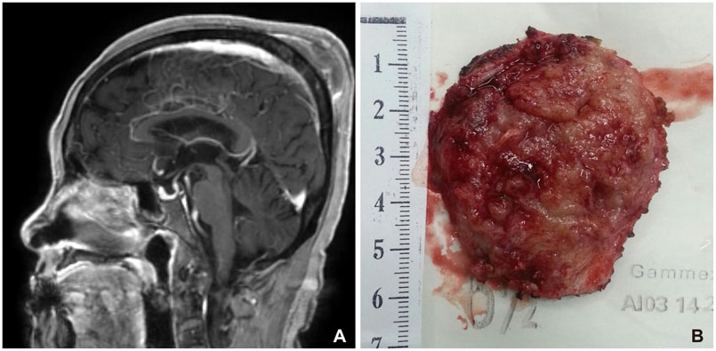
Fig. 1 A: T1-weighted, post-contrast, sagittal magnetic resonance imaging demonstrates a well-defined, heterogeneous, enhancing mass in the left parietal convexity with a focal cortical defect of the left parietal bone. B: A hard, whitish-grey mass with little bleeding was completely removed under general anesthesia.
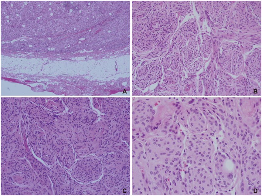
Fig. 2 Histopathologic examination revealed a non-encapsulated tumor (A: H&E, ×40) composed of nests and bundles of epithelioid to spindle cells with sparse myxoid matrix (B: H&E, ×100), arranged in fascicles or nodules, often in a geographic pattern, separated by a collagen-rich stroma (C: H&E, ×100). Mild to moderate pleomorphism of epithelioid cells with indistinct border including a giant cell and a mitotic figure is observed (D: H&E, ×200). H&E, hematoxylin and eosin.
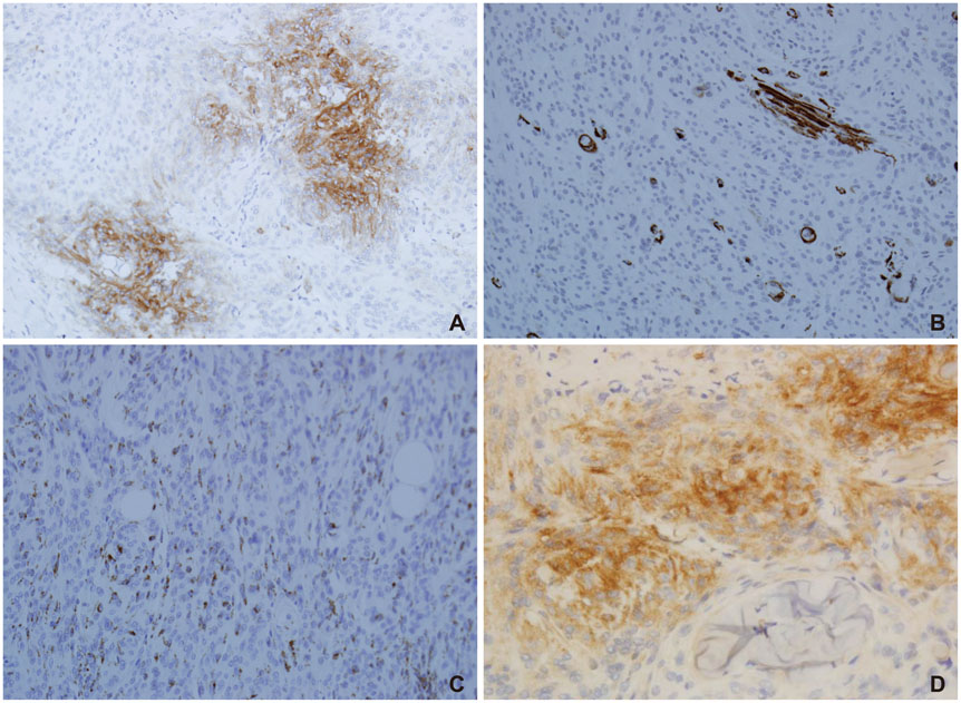
Fig. 3 Immunohistochemical studies were focally positive for vimentin, neuron-specific enolase (A: ×200), smooth muscle actin (B: ×200), CD68 (C: ×200), CD10 (D: ×200) but negative for S-100 protein, HMB-45, epithelial membrane antigen, and CD34.
Reference
-
1. Cohen NA, Samadi DS, Pawel BR, Kazahaya K. Cellular neurothekeoma of the maxilla. Ann Otol Rhinol Laryngol. 2004; 113:384–387.
Article2. D'Antonio A, Cuomo R, Angrisani B, Addesso M, Angrisani P. Cellular neurothekeoma with neuroendocrine differentiation. Dermatol Online J. 2011; 17:2.3. Pulitzer DR, Reed RJ. Nerve-sheath myxoma (perineurial myxoma). Am J Dermatopathol. 1985; 7:409–421.
Article4. Chow LT, Ma TK, Chow WH. Cellular neurothekeoma of the hypopharynx. Histopathology. 1997; 30:192–194.
Article5. Katsourakis M, Kapranos N, Papanicolaou SI, Patrikiou A. Nerve-sheath myxoma (neurothekeoma) of the oral cavity: a case report and review of the literature. J Oral Maxillofac Surg. 1996; 54:904–906.
Article6. Paulus W, Jellinger K, Perneczky G. Intraspinal neurothekeoma (nerve sheath myxoma). A report of two cases. Am J Clin Pathol. 1991; 95:511–516.
Article7. Vij M, Jaiswal S, Agrawal V, Jaiswal A, Behari S. Nerve sheath myxoma (neurothekeoma) of cerebellopontine angle: case report of a rare tumor with brief review of literature. Turk Neurosurg. 2013; 23:113–116.
Article8. Wee A, Tan CE, Raju GC. Nerve sheath myxoma of the breast. A light and electron microscopic, histochemical and immunohistochemical study. Virchows Arch A Pathol Anat Histopathol. 1989; 416:163–167.9. Hornick JL, Fletcher CD. Cellular neurothekeoma: detailed characterization in a series of 133 cases. Am J Surg Pathol. 2007; 31:329–340.
Article10. Fetsch JF, Laskin WB, Hallman JR, Lupton GP, Miettinen M. Neurothekeoma: an analysis of 178 tumors with detailed immunohistochemical data and long-term patient follow-up information. Am J Surg Pathol. 2007; 31:1103–1114.
Article11. Gallager RL, Helwig EB. Neurothekeoma--a benign cutaneous tumor of neural origin. Am J Clin Pathol. 1980; 74:759–764.
Article12. Lane JE, Mullins S, Davis LS. Clinical pathologic challenge. Cellular neurothekeoma. Am J Dermatopathol. 2006; 28:65–66. answer and discussion 87-8.13. Zedek DC, White WL, McCalmont TH. Desmoplastic cellular neurothekeoma: clinicopathological analysis of twelve cases. J Cutan Pathol. 2009; 36:1185–1190.
Article14. Remstein ED, Arndt CA, Nascimento AG. Plexiform fibrohistiocytic tumor: clinicopathologic analysis of 22 cases. Am J Surg Pathol. 1999; 23:662–670.
Article
