Histiocytic Infiltrate in Angioimmunoblastic T-Cell Lymphoma with Cutaneous Nodules and Progressive Ulceration: A Rare Manifestation
- Affiliations
-
- 1Department of Dermatology, Hallym University Sacred Heart Hospital, Anyang, Korea. dermakkh@naver.com
- KMID: 2164652
- DOI: http://doi.org/10.5021/ad.2016.28.3.391
Abstract
- No abstract available.
MeSH Terms
Figure
-
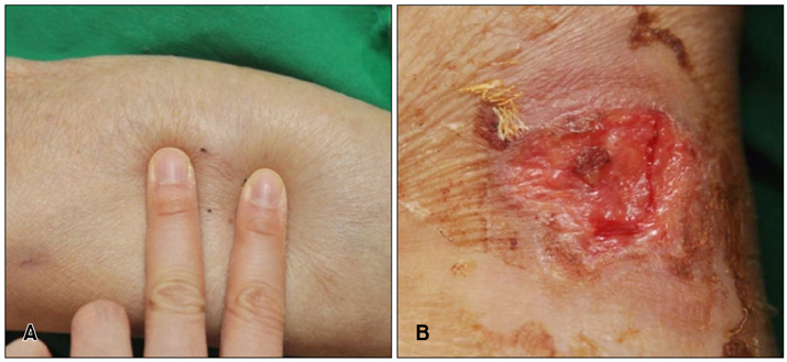
Fig. 1 (A) Localized hard subcutaneous nodules about 3.0×3.0 cm in size with no epidermal changes were palpable on both calves. (B) After several weeks, the nodule on the left calf developed into a progressive ulcer covered with granulation-like tissues, gradually penetrating into the muscle layer.
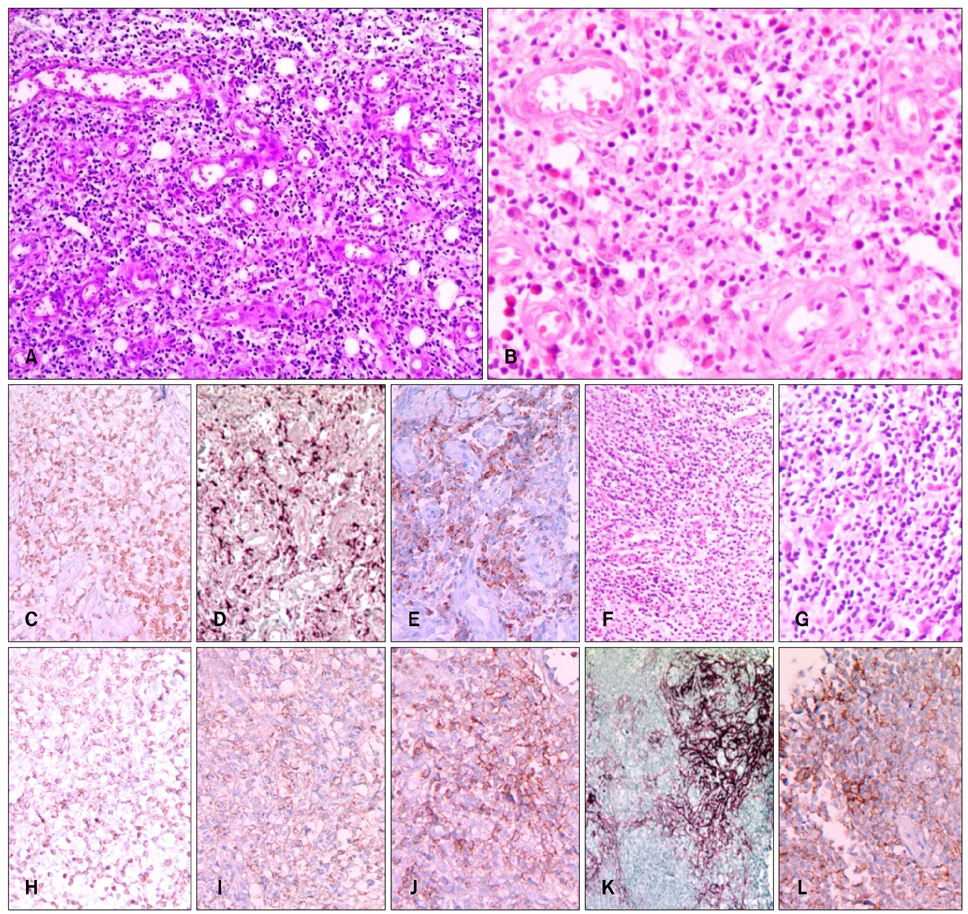
Fig. 2 (A~E) Histopathological findings of skin biopsy. (A) Dense inflammatory cell infiltration into the deep dermis (H&E, ×40). (B) Numerous small to medium-sized vascular channels surrounded by densely mixed inflammatory cells, especially abundant epitheloid histiocytes, lymphocytes, and some eosinophils (H&E, ×200). (C) Positive (CD3, ×200). (D) Strong positive (CD68, ×200). (E) Positive (programmed death-1 [PD-1], ×200). (F~L) Histopathological findings of inguinal lymph node biopsy. (F, G) Effacement of normal architecture with an interfollicular mixed polymorphous infiltrate (H&E; F: ×100, G: ×200). (H) Positive (CD3, ×200). (I) Positive (CD4, ×200). (J) Positive (CD5, ×200). (K) Irregular expansion of follicular dendritic cells (CD21, ×100). (L) Positive (PD-1, ×200).
Reference
-
1. Balaraman B, Conley JA, Sheinbein DM. Evaluation of cutaneous angioimmunoblastic T-cell lymphoma. J Am Acad Dermatol. 2011; 65:855–862.
Article2. Roncador G, Verdes-Montenegro JFG, Tedoldi S, Paterson JC, Klapper W, Ballabio E, et al. Expression of two markers of germinal center T cells (SAP and PD-1) in angioimmunoblastic T-cell lymphoma. Haematologica. 2007; 92:1059–1066.
Article3. Scarabello A, Leinweber B, Ardigó M, Rütten A, Feller AC, Kerl H, et al. Cutaneous lymphomas with prominent granulomatous reaction: a potential pitfall in the histopathologic diagnosis of cutaneous T- and B-cell lymphomas. Am J Surg Pathol. 2002; 26:1259–1268.4. Farinha P, Masoudi H, Skinnider BF, Shumansky K, Spinelli JJ, Gill K, et al. Analysis of multiple biomarkers shows that lymphoma-associated macrophage (LAM) content is an independent predictor of survival in follicular lymphoma (FL). Blood. 2005; 106:2169–2174.
Article5. Steidl C, Lee T, Shah SP, Farinha P, Han G, Nayar T, et al. Tumor-associated macrophages and survival in classic Hodgkin's lymphoma. N Engl J Med. 2010; 362:875–885.
Article
- Full Text Links
-
- Actions
-
Cited
- CITED
-
- Close
- Share
-
- Similar articles
-
- Primary Cutaneous Monomorphous Lymphoma: A Report of 3 Cases
- A case of angioimmunoblastic T cell lymphoma in a patient with ichthyosis
- A Case of Angioimmunoblastic T-cell Lymphoma Involving the Ipsilateral Parotid and Lateral Neck
- A Case of Pure Red Cell Aplasia Associated with Angioimmunoblastic T-cell Lymphoma
- A Case of Extranodall NK/T-cell Lymphoma, Nasal type
